Lobular Exophytic Gingival Swelling
Can you make the correct diagnosis?
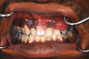
This is a 31-year-old male with an 11-month history of a sessile, lobular, progressively enlarging lesion in the anterior facial maxillary gingiva with palatal expansion.
Sorry! you are incorrect
Exostosis is a benign lamellar bony outgrowth of unknown etiology. Local mechanical irritation and genetic predisposition have been proposed as possible etiologies. They have also been described in association with free gingival grafts (1). The latter were mostly in females. They usually present in a bilateral manner, sometimes associated with maxillary or mandibular tori. Clinically, they present as protuberances of thickened cortical bone arising from the cortical plate along the buccal and posterior alveolar ridge, especially in the molar region, or in the cuspid/premolar area in the case of the free gingival graft associated exostosis (1). They are more common in the maxilla than in the mandible. They are asymptomatic unless the overlying mucosa is injured. Radiographically, they are completely radio-opaque masses and consist of histologically normal lamellar bone. Exostoses do not require treatment unless symptomatic or interfering with function. Neither the histology nor the radiographic findings in this case are supportive of exostoses.
Sorry! you are incorrect
Central ossifying fibroma is well-demarcated, sclerotic, and slow-growing, and occurs in the posterior mandible. It occurs more often in females by a ratio of 5:1, with an average age of about 35 years. However, its variant, juvenile ossifying fibroma, also known as juvenile active ossifying fibroma, does not share these characteristics (4). JOF occurs mostly in young patients, with an average age of 11 or 22 depending on the type, though reported cases range in age from six months to 70 years (2-4). It is more common in the maxilla than the mandible and has no gender predilection (3). The clinical behavior ranges from slow-growing to aggressive and locally destructive. It presents with facial asymmetry and swelling impinging on the surrounding structures. It can also occur in the frontal bone and sinuses (3-4). Those occurring in the paranasal sinuses can penetrate the nose, orbit and cranial cavity. They are usually symptomatic, presenting with pain, headache, and proptosis. Two histologic types are described: the trabecular, which occurs at an average age of 11, and psammomatoid, with an average age of 22. The psammomatoid type is more common outside the jaws (3). Radiographically, it is usually well-demarcated, but can invade the surrounding bone. It is radiolucent at the early stages and mixed radiolucent/radiopaque at more developed stages. Histologically, the trabecular type is made up of young bony trabeculae surrounded by cellular fibrous and granulation tissue stroma containing clusters of multinucleated giant cells. The psammomatoid is made up of small, rounded, cementum-like hard tissue (ossicles or psammomatoid bodies) surrounded by cellular fibrous connective tissue stroma. Treatment ranges from en bloc surgical resection to a more radical therapy reserved for the rapidly growing lesions. In spite of their invasive growth and high recurrence rate (between 30-58%) there are no reports of metastasis. The histology in this case is not supportive of juvenile ossifying fibroma.
Congratulations! You are correct
Osteosarcoma is the most common, non-hematopoietic, primary malignancy of bone. It is a malignancy of mesenchymal cells that have the ability to produce osteoid or immature bone. Osteosarcoma of the jaw represents 6-9% of all osteosarcomas. Paget’s disease and prior radiation therapy are associated with an increased risk of developing osteosarcoma. The mean age for patients with gnathic osteosarcomas is about 33 years, which is 10-15 years older than the mean age of osteosarcomas of the long bones (9-11). The maxilla and mandible are involved with equal frequency. Patients with osteosarcoma may experience pain, swelling, paresthesia and/or loosening of teeth (9-11). These tumors may vary greatly in radiographic presentation. Some lesions display an entirely radiolucent process, while others may demonstrate dense sclerosis in the affected area. However, the majority of osteosarcoma cases present as a mixed radiodense lesion. Other radiographic findings include: symmetric widening of the periodontal ligament space (PDL), diffuse borders of the lesion, periosteal reaction, “spiked” roots or the classic “sunburst” or “sun ray” appearance caused by osteophytic bone deposition at the periphery (9-11). These changes may be evident on CT, periapical, panoramic, and/or occlusal radiographic studies.
In addition to the wide range of possible radiographic presentations, the histologic findings of osteosarcoma also display a considerable amount of variability. The vast majority of conventional osteosarcomas are classified as osteoblastic, chondroblastic, or fibroblastic. The division of the specific histologic type is based on the predominant histologic pattern of the tumor. To make a diagnosis of osteosarcoma, the direct production of osteoid by malignant mesenchymal cells must be present. In addition to osteoid, the tumor cells may produce cartilaginous matrix (chondroblastic osteosarcoma) or a high-grade spindle cell stroma (fibroblastic osteosarcoma). If the majority of the tumor is composed of an osteoid product, then it is classified as osteoblastic osteosarcoma. Chondroblastic osteosarcoma is the most common histologic type seen in the jaws. The grading of osteosarcoma is based on the amount of cellular atypia; they typically graded on a scale of 1 to 4, with 4 being the highest grade (9-11).
Osteosarcoma is a persistent malignancy. Its most important prognostic indicator is the ability to obtain initial complete surgical removal. Untreated, conventional osteosarcoma is universally fatal. Osteosarcoma has an aggressive local growth potential and a propensity to spread systemically via hematogenous routes. The lung is the most frequent site of metastasis. Metastases from mandibular lesions are more frequent than from maxillary lesions. Today, therapy is typically multi-disciplinary, focusing on both local and systemic manifestations of osteosarcoma, thus incorporating surgery and chemotherapy (9-11). The use of this combined approach has resulted in a survival rate of 60-80%. In a 1997 study performed at the University of Washington, patients diagnosed with head and neck osteosarcoma had an overall 5-year survival rate of 72% (10).
Treatment
Under general anesthesia, a modified maxillectomy including teeth #s 3-13 was performed (Fig 3) and the area was replaced with a maxillary prosthesis. Many of the surgical margins were positive for osteosarcoma. Three months following the surgery, the patient presented with a mass in the maxillary sinus that was clinically suggestive of an antral mucocele caused by the original surgical intervention. However, the biopsy revealed a polypoid fragment of sinus mucosa with large and pleomorphic cells consistent with osteosarcoma. The patient is currently undergoing proton therapy.
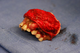
Figure 3. This represents the surgical specimen from the modified maxillectomy procedure. It includes teeth #s 3-13. The area is replaced with a maxillary prosthesis.
References
- Echeverria JJ, Montero M et al. Exostosis following a free gingival graft. J Clin Periodontol. 2002;29:474-477.
- Rinaggio J, Land M, Cleveland DB. Juvenile ossifying fibroma of the mandible. J Pediatr Surg. 2003;38:648-650.
- El-Mofty S. Psammomatoid and trabecular juvenile ossifying fibroma of the craniofacial skeleton: two distinct clinicopathologic entities. Oral Surg Oral Med Oral Pathol Oral Radiol Endod. 2002;93:296-304.
- MacDonald-Jankowski DS. Fibro-osseous lesions of the face and jaws. Clin Radiol. 2004;59:11-25
- Rao VV, Schnittger S et al. G protein Gs alpha (GNAS 1), the probable candidate gene for Albright hereditary osteodystrophy, is assigned to human chromosome 20q12-q13.2. Genomics. 1991;10:257-261
- Neville BW. Oral & maxillofacial pathology. 2nd ed. Philadelphia: W.B. Saunders; 2002
- Anil S, Beena VT et al. Chondrosarcoma of the maxilla. Case report. Aust Dent J. 1998;43:172-174.
- Hayt MW, Becker L et al. Chondrosarcoma of the maxilla: panoramic radiographic and computed tomographic with multiplanar reconstruction findings. Dentomaxillofac Radiol. 1998;27:113-116.
- Bennett JH, Thomas G et al. Osteosarcoma of the jaws: a 30-year retrospective review. Oral Surg Oral Med Oral Pathol Oral Radiol Endod 2000;90:323-332
- Oda D, Bavisotto LM et al. Head and neck osteosarcoma at the University of Washington. Head Neck 1997;19:513-523.
- Slootweg PJ, Muller H. Osteosarcoma of the jaw bones. Analysis of 18 cases. J Maxillofac Surg 1985;13:158-166.
Sorry! you are incorrect
This is a malignant mesenchymal neoplasm of cartilage origin. It is uncommon, especially in the jaws. However, it is still much more common than the benign counterpart (chondroma) in the jaws. It occurs more often in the maxilla, particularly in the incisor area. It can occur at any age but is most common in males in the sixth decade of life (7-8). Unlike osteosarcoma, it has a low tendency for metastasis. In general, the prognosis is better than that of osteosarcoma, but can vary depending on the stage of the disease. It commonly presents as an asymptomatic swelling with buccal and lingual expansion. Patients may experience unexplained paresthesia, headache, loosening and loss of teeth. Radiographically, it presents as an ill-defined, mottled radiolucency with snowflake or punctate calcifications (7-8). Sometimes, the teeth will show a widened periodontal ligament space. Histologically, it is characterized by immature and pleomorphic cartilage, but at times the cartilage is benign-looking. It is rare for the cartilage to calcify or differentiate and form bone (7-8). Therapy for chondrosarcoma ranges from wide local excision to radical resection with or without chemotherapy, depending on the stage of disease. The prognosis ranges from good to poor. The histology in this case is not supportive of chondrosarcoma.
Sorry! you are incorrect
Fibrous dysplasia is a tumor-like developmental and hamartomatous disorder of bone that presents in three forms: monostotic, polyostotic and craniofacial. It is common, affecting 7% of all benign bone tumors. The monostotic form constitutes approximately 80% of all fibrous dysplasia cases. It affects males and females equally, occurs in childhood and adolescence and usually completes growth by the age of 30. The craniofacial form affects the maxilla and the craniofacial complex, and is more aggressive than the monostotic form. Polyostotic cases constitute 10% of all FDs; they may (such as McCune-Albright) or may not (Jaffe) be associated with multiple endocrine disorders (4, 6). The etiology of FD is not clear but a mutation (GNAS 1) has been identified at multiple chromosomes. This gene can occur at all ages, even in infancy (5). Some of the FD cases are clinically asymptomatic and are found incidentally; many present with swelling and deformity, and rare cases involve pain. Swelling of the maxilla or mandible is the most common presentation. It may involve bones other than the maxilla, including the zygoma, sphenoid and frontal. Monostotic FD is usually unilateral and is known to displace the surrounding teeth, but is otherwise firmly seated. It is usually slow-growing, but rapid growth has been described, especially during puberty. With rapid growth, temporary pain is also described. The radiographic appearance, especially of the maxilla, is classically described as a ground glass appearance where fine radiopacity is noted (4-6). The mandibular lesions are much more deceiving; more variability exists, thus making the radiographic diagnosis more difficult. It ranges from cystic radiolucency to a classical ground glass radio-opacity. Histologically, it consists of vascular fibrous connective tissue stroma with irregular (Chinese character) spicules of woven bone. Giant cells may be seen. At times, artifactual shrinkage of the connective tissue from bone is noted. Many cases do not need treatment; those that do are best treated with surgical recontouring of the affected bone to address aesthetic or functional concerns (4-6). This is preferably performed after cessation of growth due to the high incidence of requirement for secondary procedures. Some sources report a tendency for increased growth of the lesion following surgical intervention, but the evidence for this is weak. Even if recontouring is performed in adulthood, multiple procedures are frequently required. This may be due to inadequate removal, continued growth or the ossification of the subperiosteal hematoma, which is difficult to avoid due to the very vascular nature of the dysplastic bone. Radical excision with primary reconstruction of the affected bone has been suggested, but is not widely accepted. Radiation therapy has been used in the past and is successful in stopping growth of the lesion. Unfortunately, a significant incidence of development of osteosarcoma in the irradiated bone prohibits the use of this modality. Treatment with bisphosphonates has also been successfully used. The overall prognosis is good with proper clinical management. The age of the patient, the histology and the radiographic findings are supportive of a diagnosis of FD.