Well-defined radiopaque lesion right posterior mandible
Contributed by Dr. Libby Kutcipal
Ballard Oral & Maxillofacial Surgery, Seattle, WA
Case Summary and Diagnostic Information
This is a 14-year-old male with an otherwise asymptomatic lesion discovered by the patient’s general dentist during a routine dental check-up.
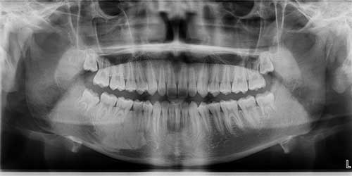
This is a 14-year-old male with an otherwise asymptomatic lesion discovered by the patient’s general dentist during a routine dental check-up. On panoramic imaging, a large and well-defined homogenous radiopaque lesion was identified between teeth #s 28-31 extending near the inferior border of the mandible (Figure 1). The area was otherwise not painful, and teeth showed no evidence of resorption or displacement. Expansion was mild but not clinically significant.
Figure 1 Panoramic radiograph taken at routine dental visit. Note the diffuse, well-defined homogeneous radiopaque (RO) lesion between teeth #s 28 and 31. The teeth are not displaced and show no evidence of resorb.
The patient’s past medical history is negative.
The patient presented to the Oral Surgery clinic with the following complaint: “My dentist said I have an area on the lower right side jaw that does not look normal.” There were no clinical symptoms and the duration of the lesion was unknown (Figure 1). The teeth showed no evidence of resorption or displacement. The area was biopsied.
A small incisional biopsy was performed under local anesthesia and N2O/O2 sedation. The specimen was submitted for microscopic evaluation.
Histologic examination of the decalcified and H & E stained section revealed small fragments of lamellar bone with viable osteocytes (Figures 2 & 3).
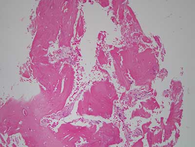
Figure 2 Low power (X100) histology shows H & E stained section with small fragments of decalcified bone and small fragments of connective tissue. Bone comprises the bulk of the specimen and is mostly lamellar in type. It is made up of Haversian system containing viable osteocytes. Inflammatory cells are absent.
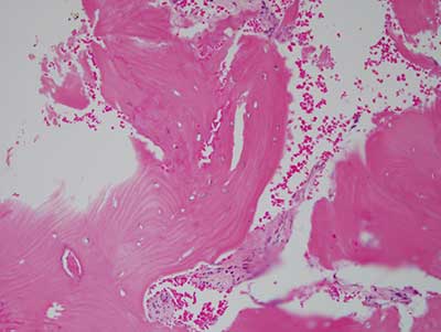
Figure 3 Higher power (X200) histology shows H & E stained section with closer look at the decalcified lamellar bone with viable osteocytes.
After you have finished reviewing the available diagnostic information