Large mixed radiolucent radiopaque lesion,
area of impacted tooth # 32
Can you make the correct diagnosis?
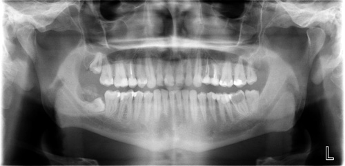
This is a 29-year-old white male referred by his general dentist for a swelling distal to tooth # 32. It was of unknown duration and the patient reported pain in the area. The size of the lesion was about 4 cm at its greatest dimensions. Panoramic radiograph showed a well-demarcated lesion of mixed bony origin with a radiopaque mass in the center and a prominent radiolucent rim at the periphery (Figure 1). Although closely associated, this lesion was clinically separate from the impacted tooth #32.
Sorry! you are incorrect
If we interpret the radiographic findings are a lesion clearly associated with impacted tooth #32, then our differential diagnosis should only focus on conditions that are of tooth origin and are capable of producing hard tissue such as odontoma, calcifying odontogenic cyst, ameloblastic fibro-odontoma, calcifying epithelial odontogenic tumor and others. For the sake of time, I will cover Odontoma and COC since our patient is on the older age range for ameloblastic fibro-odontoma.
The radiographic findings and the appearance of close association with tooth # 32 are supportive of odontoma. The histology however is not.
Odontoma, is the most common odontogenic tumor, accounting for 22% of all odontogenic tumors. It is of mixed epithelial and mesenchymal origin. It is usually associated with unerupted teeth (1). It can occur at any age, but is more common in the first two decades with an average age of 14 or 18. It is slightly more common in females and more common in the maxilla, especially the anterior maxilla, than in the mandible. Compound odontomas are more common in the anterior jaws while complex odontomas are more common in the posterior jaws. About 80% of odontomas are associated with impacted teeth. Radiographically, odontomas present as well-circumscribed radiolucency resembling a dental follicle or dentigerous cyst with one or multiple radiopaque pieces resembling teeth. Compound odontomas tend to occur between teeth and tend to be composed of multiple small tooth-like structures while complex odontomas tend to occur in the posterior jaws and present as a conglomerate mass. Both types are made up of enamel matrix, dentin, cementum, and dental pulp surrounded by a dental follicle or cyst. Histologically, the tooth-like structures are arranged in a uniform manner similar to the normal tooth. These lesions are benign and are conservatively treated with simple curettage (2). Recurrence is rare and if it recurs, one must rule out other odontogenic lesions such as COC, ameloblastic fibro-odontoma and others.
Sorry! you are incorrect
The radiographic findings and the appearance of close association with tooth # 32 are also supportive of calcifying odontogenic cyst associated with odontoma. The histology however is not.
Calcifying odontogenic cyst is not a common condition, accounting for only 1% of jaw cysts. This condition represents a spectrum of histologies including a simple cyst, a cyst with an odontoma, a cyst with ameloblastomatous proliferations and a more aggressive solid neoplasm, also known as “ghost cell odontogenic tumor,” with a potential to transform especially with multiple recurrences. It is therefore important to render a specific histological diagnosis. Calcifying odontogenic cyst is also described on the gingiva alone without a bony component in up to 20% of cases (peripheral COC). Intrabony COC presents as a well-circumscribed unilocular radiolucency with flecks or masses of radiopaque material. The size and amount of the radiopaque material varies depending on the type of cyst and whether or not it is associated with an odontoma. It can expand bone, displace and resorb teeth. In about one third of cases, COC is associated with unerupted teeth, usually the canine tooth. The most common locations are the anterior jaws in the incisor-canine area. It can occur at any age, but is most common in patients around 33 years of age (3-4); however, the odontoma-associated COCs tend to occur in younger females around the age of 17. This cyst may also be associated with an odontoma, usually complex in type. The solid odontogenic ghost cell tumor is the least common, but behaves more aggressively than the others. These lesions are usually conservatively treated by thorough curettage and heal uneventfully (4). The solid type tends to recur and, for that reason, follow-up visits are recommended. With recurrence, a more aggressive treatment is recommended to prevent further recurrence.
Sorry! you are incorrect
If we take the clinical finding of this lesion being clearly separate from the impacted tooth #32, we should at this point expand the differential diagnosis to include non-odontogenic, bone forming conditions such as central ossifying fibroma, osteoid osteoma and osteoblastoma.
The radiographic presentation is supportive of a mature COF, the histology is not supportive of this condition. In addition, the far posterior mandible (distal to tooth #32) is unusual location for COF.
Central ossifying fibroma is a benign neoplasm of bone origin. It presents as a well-demarcated to corticated radiolucent or mixed radiolucent/radiopaque mass with a peripheral radiolucent rim (5-6). Central ossifying fibroma is a slow-growing, expansile lesion with characteristic downward expansion of the inferior border of the mandible. It can also expand buccally and lingually. The associated teeth are vital. It is common in young adults around 35 years of age and is five times more likely to occur in females than males. It affects the posterior mandible in about 90% of cases (5-6).
Congratulations! You are correct
Osteoblastoma is a very rare benign neoplasm of osteoblast origin. It is similar histologically, radiographically and clinically to osteoid osteoma but larger in size. for that reason, osteoblastoma was at one point referred to as “giant osteoid osteoma” (7). Osteoid osteomas tend to be smaller than 2cm in diameter while osteoblastomas are larger than 2 cm. This patient’s lesion is described as 4 cm in size. Osteoblastomas are usually slow-growing but can occasionally be aggressive and thus the variant of aggressive osteoblastoma with features that can be misinterpreted as osteosarcoma (8-10). Aggressive osteoblastomas tend to be larger than 4 cm and tend to occur in individuals older than 30 years of age (9). They too are associated with pain (9).
Osteoblastomas are rare in general and certainly very rare in the jaw bone (9-10). They are most common in the vertebral column and long bones. In the vertebrae, they may cause spinal cord or nerve compression. They are also described in the calvarium, small bone of the hand and feet and sacral bone as well as the jaws especially the posterior mandible (8-10). Clinically, they occur in individuals under 30 years of age with slight predilection to females. They are associated with dull pain which is less responsive to Aspirin compared to pain associated with osteoid osteomas where patients often get relieve with Aspirin. Osteoblastomas are expansile and can displace and resorb teeth.
Radiographically, osteoblastomas are more often well-demarcated mixed radiolucent/radiopaque mass with variable degree of mineralization. Occasionally osteoblastomas may have an ill-defined border.
Histologically, osteoblastomas demonstrate a combination of young bone, active osteoblasts, plump osteocytes in large lacunae, osteoclasts and vascular connective tissue stroma. The bony trabeculae are of variable shapes and sizes with reversal lines. The bony trabeculae are lined by many osteoblasts and osteoclasts. The connective tissue supporting the hard tissue is usually loose and vascular with many congested blood vessels. The osteoblasts are large but mature and show no evidence of atypia. The mineralized bone is also mature.
Complete surgical excision is the treatment of choice. Since this case is categorized as non-aggressive, treatment would be thorough curettage as was done. For the aggressive type, resection would be a more appropriate treatment. The overall prognosis is good and recurrence rate is uncommon for the conventional osteoblastoma while significantly higher for the aggressive type.
References
- Yeung KH, Cheung RC et al. Compound odontoma associated with an unerupted and dilacerated maxillary primary central incisor in a young patient. Int J Paediatr Dent. 2003;13:208-212.
- Chang JY, Wang JT et al. Odontoma: a clinicopathologic study of 81 cases. J Formos Med Assoc. 2003;102:876-882.
- Gorlin RJ, Pindborg JJ et al. The calcifying odontogenic cyst–a possible analogue of the cutaneous calcifying epithelioma of Malherbe. An analysis of fifteen cases. Oral Surg Oral Med Oral Pathol. 1962;15:1235-1243.
- Moleri AB, Moreira LC et al. Comparative morphology of 7 new cases of calcifying odontogenic cysts. J Oral Maxillofac Surg. 2002;60:689-696.
- Su L, Weathers DR, et al. Distinguishing features of focal cemento-osseous dysplasia and cemento-ossifying fibromas: II. A clinical and radiologic spectrum of 316 cases. Oral Surg Oral Med Oral Pathol Oral Radiol Endod 1997 Nov; 84: 540-549.
- Chang CC, Hung HY, Chang JY, Yu CH, Wang YP, Liu BY, Chiang CP. Central ossifying fibroma: a clinicopathologic study of 28 cases. J Formos Med Assoc. 2008 Apr;107(4):288-94.
- Dahlin D, Johnson E. Giant Osteoid Osteoma. J Bone Joint Surg. 1954;36-A:559-572.
- Biagini R, Orsini U, Demitri S. Osteoid osteoma and osteoblastoma of the sacrum. Orthopedics. Nov 2001;24(11):1061-4.
- Lypka MA, Goos RR, Yamashita DD, Melrose R. Aggressive osteoblastoma of the mandible. Int J Oral Maxillofac Surg. 2008 Jul;37(7):675-8.
- Jones AC, Prihoda TJ, Kacher JE, Odingo NA, Freedman PD. Osteoblastoma of the maxilla and mandible: a report of 24 cases, review of the literature, and discussion of its relationship to osteoid osteoma of the jaws. Oral Surg Oral Med Oral Pathol Oral Radiol Endod. 2006 Nov;102(5):639-50.