Exophytic yellow nodule; right posterior mandibular vestibule
Can you make the correct diagnosis?
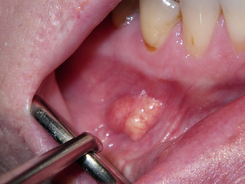
This is a 54-year-old white female who presented with an exophytic, smooth-surfaced yellow nodule in the posterior right mandibular vestibule of unknown duration.
Sorry! you are incorrect
The color, the exophytic appearance; the soft consistency and the age of the patient are all in support of the clinical diagnosis of a lipoma. The vestibule, however is a rare location for a lipoma; it occurs more often in the buccal mucosa than in the vestibule. The histology is not supportive of a lipoma.
Lipomas are benign neoplasms of adipose tissue origin. They are more commonly described in the trunk and extremities and are rare in the oral cavity. They constitute around 4.4% of all benign oral lesions. In a recent study from the Armed Forces Institute of Pathology (AFIP) of 125 benign lipomas in and around the mouth, the male to female ratio was approximately 3:1, which is not surprising, given the study was extracted from a military population. Other studies reveal an equal gender distribution. The AFIP study showed the mean age to be 52 with a range of 9-92 years. Only 4 of the 125 cases involved patients under 18. In the mouth, the most common location for this neoplasm is the buccal mucosa, followed by the lips, submandibular area, tongue, palate and, less frequently, on the floor of mouth and vestibule. These lesions are slow-growing and are usually of long durations. Lipomatous nodule of the buccal mucosa may represent herniation of the buccal fat pad. Lipomas usually present as a single, smooth-surfaced, soft (with a doughy consistency), lobulated, painless, yellowish, and sessile nodule. The overlying mucosa is usually thin and stretched with visible blood vessels. Because of its softness it can be mistaken for a cyst. Histologically, lipomas are variable benign histologies. Some have predominant lobules of mature adipocytes surrounded by a thin connective tissue capsule, while others have a predominant spindle-cell component, or a myxoid, chondroid connective tissue component. Some lipomas occur intramuscularly. Each has its own clinical behavior. Simple surgical excision is the treatment of choice for the simple, mature adipocyte component-type lipomas.
Sorry! you are incorrect
Although keratin-filled cysts, especially superficial cysts, would reflect as white in color, they are sometimes also yellow in color. Therefore, the yellow color, the exophytic appearance and the soft consistency is supportive of the clinical diagnosis of a keratin-containing cyst such as an epidermal cyst or a benign lymphoepithelial cyst. However, the vestibule is a rare location for either of these two cysts. The histology is not supportive of either of the two cysts.
Benign lymphoepithelial cyst (BLC) is a developmental cyst of the lateral neck and the oral cavity. The lateral neck is also known as the branchial cleft or cervical lymphoepithelial cyst. The oral cavity counterpart is known as the benign lymphoepithelial cyst. The names come from the main histologic components of a cystic structure lined by keratinized epithelia and connective tissue with aggregates of lymphocytes. The origin is from epithelium entrapped in lymphoid aggregates. In the oral cavity, it presents as a small yellowish-white raised lesion most commonly occurring in the floor of mouth, ventral tongue and tonsillar area. The vestibule is not a common location for BLC. It is histologically made up of a cystic cavity lined by keratinized epithelium and supported by fibrous connective tissue containing many lymphocytes. The latter are mature, some form follicles with germinal centers. Conservative surgical excision is the treatment of choice.
Epidermal cyst also called epidermoid cyst or epidermal inclusion cyst, is common on the skin but is rare in the oral cavity. On the skin, it is sometimes mistakenly called sebaceous cyst. A cystic structure similar to epidermoid cyst occurs in the midline floor of mouth and is known as dermoid cyst. Dermoid cyst is believed to be a teratoma such as that described in the ovary, but less differentiated. Epidermal cyst is believed to be an entrapment of surface epithelium in the underlying connective tissue. Multiple skin epidermoid cysts should make a clinician consider a diagnosis of Gardner’s syndrome. Epidermal cyst presents as nodular and fluctuant subcutaneous swelling. It is usually asymptomatic, but can be painful if inflamed. If superficial, it may be white or yellow in color. The lips and tongue are the most common locations for this cyst. It is rare in the vestibules. Epidermal cyst is made up of a cystic structure lined by keratinized epithelium and supported by dense connective tissue. The lumen is filled with keratin. Conservative surgical removal is the treatment of choice.
Sorry! you are incorrect
The age and gender are good for verruciform xanthoma (VX). However, the yellow color is the main reason why VX is included on the differential diagnosis. The smooth-surface exophytic appearance and the location are unusual for VX. Although VX is sometimes exophytic, the surface is usually rough and warty. The histology is not supportive of VX.
Verruciform xanthoma is a benign condition of unknown etiology and pathogenesis but is favored to be an inflammatory process rather than a neoplasm. A viral etiology has been suggested, especially in association with human papilloma virus. It has been described in connection with oral lichen planus, recessive dystrophic epidermolysis bullosa, graft vs. host disease, congenital hemidysplasia with ichthyosis for erythroderma, limb defect (CHILD) syndrome, and epidermal nevus.
VX was first described in the oral cavity by Shafer et al in 1971, and has since been reported in many other areas, including the anogenital area and skin. VX usually presents as a solitary lesion but can also present in multiples. It ranges in diameter from a few millimeters to two centimeters (cm) with an average size of 1cm. However, larger VXs several cm in size have been described. They can be flat or raised and warty and can be reddish-pink, yellow, orange, white or gray. VX is most commonly described in patients in the fifth and sixth decade of life with a 2:1 predilection for occurrence in females. VX of the scrotal area is described mostly in older Japanese males with a wide age range. It is rare in children in general. It is usually asymptomatic and does not have a specific clinical presentation. However, when presenting as yellowish or orange-like in color, it can be clinically diagnostic. It can occur anywhere in the mouth, but the gingiva and the alveolar ridge constitute 50% of all cases, followed by the palate. The vestibule is a rare location for VX.
The histology is characterized by papillary epithelium with evenly aligned rete pegs. The papillary projections are short, extending slightly above the surface. The projections have deep clefts filled with keratin. They are supported by connective tissue papillae, which are densely packed with large cells with foamy cytoplasm that are referred to as xanthoma cells. The latter are usually in the connective tissue papillae, but can extend into the surface epithelium and into the connective tissue below the superficial papillae. The xanthoma cells are believed to be of monocyte-macrophage lineage. Inflammatory cells and microabscesses can be present within the epithelium as well as the connective tissue. Treatment consists of simple excision and recurrence is rare.
Congratulations! You are correct
Cystadenoma is a rare and unusual entity which may represent a benign neoplasm or a reactive process depending on whose research one reads. Those who characterize it as a benign neoplasm suggest that it is adenomatous in origin and affects a variety of tissues including minor and major salivary gland tissues, the ovaries, epididymis, pancreas and other sites. The neoplastic lesions are typically described to be multicystic with papillary proliferations within the cystic structures but can also, less often, be unicystic. Those who believe it to be a reactive condition such as intraductal papillary hyperplasia or associated with an obstructive process such as mucous retention cyst (salivary duct cyst) with papillary proliferations and at times with combined oncocytic metaplasia, mucous producing cells and papillary proliferations as is the case with our patient. Whether the histology of this case is that of a reactive process or a true neoplastic process is open to debate, but what is clear is that it is benign and has a multicystic component with focal areas of oncocytic metaplasia and papillary proliferation all support a diagnosis of papillary cystadenoma. It however lacks the solid tumor component which would raise the question of whether this is an obstructive reactive process with papillary hyperplasia and oncocytic metaplasia with mucous cells.
Cystadenomas of the major and minor salivary gland tissue are more common in females by a 2:1 ratio around 55 years of age. In the oral cavity, it is described to occur on the lips, palate and buccal mucosa. This case was in the vestibule, which is close to the buccal mucosa area. Cystadenomas are clinically described to be similar to a mucocele, which is usually light blue to pink depending on the depth of the lesion. The yellow color is highly unusual for a mucocele or a cystadenoma, especially when it is not infected. Yellow usually indicates anything like pus, keratin, fat and/or sialolith rather than a mucous-filled cavity. Cystadenomas tend to be slow-growing and asymptomatic.
Histologically, cystadenomas are usually multicystic with a solid component within the surrounding connective tissue and papillary projections in the lumen of the cystic structures. They can also be unicystic with a solid-tumor component and papillary projections. Because the papillary projections are frequently present, “papillary cystadenomas” is more commonly used as the name of this condition. These lesions can have a prominent mucous cell component or an oncocyte component, hence the names “papillary oncocytic cystadenoma” and “papillary mucous cystadenoma.” The histologic differential diagnosis includes low-grade mucoepidermoid carcinoma, papillary cystic acinic cell carcinoma, sclerosing polycystic adenosis, salivary duct cyst with papillary hyperplasia and low-grade cystadenocarcinoma. Our case is clearly well-defined and benign with no evidence of invasion.
Conservative surgical removal is the treatment of choice; these lesions rarely recur.
References
- Furlong MA. Fanburg-Smith JC, Childers ELB. Lipoma of the oral and maxillofacial region: Site and subclassification of 125 cases. Oral Surg Oral Med Oral Pathol Oral Radiol Endod. 2004 Oct;98(4):441-50.
- Fregnani E.R, Pires FR, Falzoni R, Lopes MA and Vargas PA. Lipomas of the oral cavity: clinical findings, histological classification and proliferative activity of 46 cases. Int J Oral Maxillofac Surg 32 (2003), pp. 49–53.
- Fletcher C.D.M. Unni KK and Mertens F. Adipocytic tumors. In: Pathology and genetics: tumours of soft tissue and bone. World Health Organization classification of tumours, IARCPress, Lyon, France (2007), pp. 9–46.
- Khelemsky R, Mandel L. Lymphoepithelial cyst of mouth floor. J Oral Maxillofac Surg. 2010 Dec;68(12):3055-7.
- Gallagher G, Kabani S, Noonan V. Oral lymphoepithelial cyst. J Mass Dent Soc. 2006 Winter;54(4):52.
- Ozan F, Polat HB, Ay S, Goze F. Epidermoid cyst of the buccal mucosa: a case report. J Contemp Dent Pract. 2007 Mar 1;8(3):90-6.
- Jham BC, Duraes GV, Jham AC, Santos CR. Epidermoid cyst of the floor of the mouth: a case report. J Can Dent Assoc. 2007 Jul-Aug;73(6):525-8.
- Ide F, Obara K, Yamada H, Mishima K, Saito I, Kusama K. Cellular basis of verruciform xanthoma: immunohistochemical and ultrastructural characterization. Oral Dis. 2008 Mar;14(2):150-7.
- Philipsen HP, Reichart PA, Takata T, Ogawa I (2003). Verruciform xanthoma – biological profile of 282 oral lesions based on a literature survey with nine new cases from Japan. Oral Oncol 39: 325–336.
- Polonowita AD, Firth NA, Rich AM (1999). Verruciform xanthoma and concomitant lichen planus of the oral mucosa. A report of three cases. Int J Oral Maxillofac Surg 28: 62–66.
- Cheu KW, Chan JKC. Salivary gland tumors (2007). Fletcher C.D.M. Diagnostic histopathology of tumors. Third edition, Churchill Livingstone Elsevier. pp 239-325.
- Auclair PL, Ellis GL, Gnepp DR. Other benign epithelial neoplasms. Ellis GL, Auclair PL and Gnepp DR (1991). Surgical pathology of the salivary glands. Philadelphia, PA, W.B Saunders. pp 252-268.
- Waldron CA, El-Mofty SK, Gnepp DR. Tumors of the intraoral minor salivary glands: a demographic and histologic study of 426 cases. Oral Surg Oral Med Oral Pathol. 1988 Sep;66(3):323-33.
- Mahler V, Schell H. Papillary cystadenoma: a rare tumor of the minor salivary glands. Eur J Dermatol. 1999 Jul-Aug;9(5):387-9.