Well-defined radiopaque lesion right posterior mandible
Can you make the correct diagnosis?
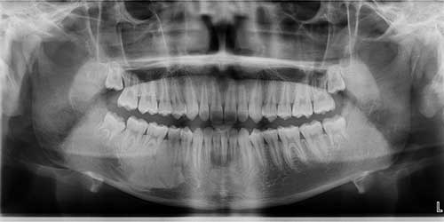
This is a 14-year-old male with an otherwise asymptomatic lesion discovered by the patient’s general dentist during a routine dental check-up.
1. Fibrous dysplasia
2. Chronic sclerosing osteomyelitis
3. Benign cementoblastoma
4. Idiopathic osteosclerosis
Sorry, you are incorrect!
A differential diagnosis for a homogeneous radiopaque lesion of the jaws should include fibrous dysplasia, especially when the patient is a child. The age and the radiopaque nature of this lesion is consistent with FD. However, the site, the well-defined borders, and the lack of expansion are not consistent with fibrous dysplasia. The homogenous look of the radiopacity is also not typical of FD since this condition tends to have more of a ground glass or cotton wool appearance.
That said, FD of the mandible can present in atypical manner compared to that of the maxilla and can be well-defined rather than blending in with the surrounding normal bone. For that reason, we should include it on the differential diagnosis. However, the histology is not consistent with FD.
Sorry, you are incorrect!
It is unusual for chronic sclerosing osteomyelitis (CSO) to occur in children but it has been reported under the name “juvenile mandibular chronic osteomyelitis” and similar titles. The age and site in this patient are consistent with that described in CSO. The gender is not consistent with most cases of CSO. The female-to-male ratio of CSO is 3:1. The clinical presentation is also not consistent with CSO; in children and adults alike, it is usually painful, and the pain intensifies periodically. In children, CSO is also associated with soft tissue swelling of the involved site. In this case, the lesion was discovered as incidental finding. Another aspect of the clinical presentation in this case that is not typical of SCO is the lack of significant swelling. The radiographic findings are also not consistent with chronic sclerosing osteomyelitis since the latter tends to be irregular and mixed RL/RO (although predominantly RO). It also tends to be diffuse with ill-defined borders. Finally, the histology is not consistent with chronic sclerosing osteomyelitis.
Sorry, you are incorrect!
The age, the gender, and the site in this case are all consistent with benign cementoblastoma. However, the lack of clinical symptoms, the involvement of multiple teeth and the radiographic findings are not. Benign cementoblastomas tend to affects one tooth rather than multiple teeth, as is the case in this patient. Benign cementoblastomas occur more often in young males under the age of 25, sometimes with pain, and they are usually expansile. They usually affect the mandibular first molar but also the second molar. They are usually apical but can extend laterally. They usually resorb and fuse to the root(s) of the tooth. Radiographically, benign cementoblastoma is usually mixed RL/RO and is surrounded by a rim of radiolucency with an outer corticated border. The histology in this case is not consistent with benign cementoblastoma.
Congratulations, you are correct!
The site and radiographic finding of homogeneous radiopacity is typical of idiopathic osteosclerosis (IO). These lesions can occur at any age. Nearly 60% of cases occur in females. Idiopathic osteosclerosis is usually asymptomatic and tends to be discovered as an incidental finding, which is the case in this patient. These lesions are usually small and round-to-oval in shape. They tend present apical to a single tooth (premolar or molar mandibular teeth), away from teeth; this is not consistent with this patient’s presentation of a lesion involving multiple teeth and extending between teeth. Only 2% of IO cases involve more than one tooth. Close to 20% involve the apex of teeth and extend between teeth, which is the case with this patient. The histology in this case is consistent with the diagnosis of idiopathic osteosclerosis.