Large mixed radiolucent/radiopaque mass: Left maxilla & maxillary sinus
Can you make the correct diagnosis?
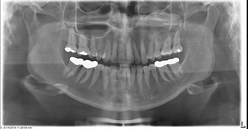
This is a 59-year-old female who noted a swelling in her left maxilla for a few months that she described as slow growing and painful. Tooth mobility in the vicinity of the swelling was the main source of pain.
Sorry, you are incorrect!
The site in the maxilla and the mixed radiolucent-radiopaque mass filling the maxillary sinus are clinical and radiographic features consistent with fibrous dysplasia (FD). The lack of a ground-glass appearance makes a diagnosis of FD less likely, as does the older age of this patient. Over 85% of FD cases occur in the first two decades of life, and they are rare after age 30. In addition, the radiographic presentation of a well-demarcated superior aspect is not consistent with FD since these lesions are usually diffuse and blend in with the surrounding normal bone. The pain and tooth mobility are also not consistent with the behavior of craniofacial monostotic FD. The histology in this case is not consistent with fibrous dysplasia.
Were this lesion FD, it would be a monostotic (occurring in one bone or bone complex area) FD. This type constitutes approximately 80% of all fibrous dysplasia cases. FDs of the craniofacial area are expansile and disfiguring lesions, but they are not painful. Monostotic FD in the jaws affects males and females equally. It typically occurs in the first two decades of life, more often during childhood and puberty, and usually stops growing by age 30. It appears as an asymptomatic swelling of the maxilla or, less often, the mandible. It may involve bones other than the maxilla, including the zygoma and sphenoid. It is usually unilateral and can displace teeth but it does not cause tooth mobility. FD usually grows slowly but rapid growth has been described, especially during puberty. The radiographic appearance of FD, especially when occurring in the maxilla, is typically of ground glass where fine radiopacity is noted. Mandibular FD lesions are much more deceptive because they tend to vary in radiographic appearance from cystic unilocular radiolucency to multilocular radiolucency to the classic ground glass radio-opacity.
Sorry, you are incorrect!
The site in the maxilla, the age and gender of this patient, and the radiographic findings of a mixed radiolucent/radiopaque lesion are all features consistent with the clinical and radiographic behavior of central odontogenic fibroma. The well-demarcated superior portion of the lesion is also consistent with central odontogenic fibroma, as is the tooth mobility. The histology is not consistent with central odontogenic fibroma.
Central odontogenic fibroma is a rare neoplasm of mesenchymal odontogenic origin. Histologically, it is madeup of connective tissue stroma with odontogenic epithelial nests and may or may not have calcified material simulating cementum globules. Central odontogenic fibroma occurs in a wide age range of 9-80 with a mean patient age of 40 years. It occurs more commonly in females with a ratio of 2:1 or 7:1, depending on the studies reported. About 60% of cases occur in the anterior maxilla and the rest in the posterior mandible, usually between teeth, though about one third are associated with impacted teeth. Some of the neoplasms that occur in the maxilla can cause palatal depression (dimpling) of the hard palate below where the neoplasm is. These neoplasms are slow growing but can reach large sizes. They can resorb and displace teeth as well as cause tooth mobility. Radiographically, they can be unilocular or multilocular, expansile, and completely radiolucent. In about 12% of cases, they can be radiolucent with flecks of radiopacity.
Congratulations, you are correct!
The site of this case makes a diagnosis of CGCG less likely because 70% of CGCG cases occur in the mandible anterior to the first molar and, at times, crossing the midline. The mixed radiolucent/radiopaque radiographic presentation is also not typical for this condition because the majority of cases present as multinodular radiolucent lesions. In addition, the age of this patient is not typical of CGCG cases; about 60% of cases occur under in patients the age of 30. The gender of the patient is consistent with CGCG since this lesion affects females more often than males with a female-to-male ratio of 2:1. The histology is that of CGCG producing bone. The bone formation can account for the mixed RL/RO radiographic findings. This lesion is recurrent by history. It was first diagnosed and treated surgically in 2016 and recurred in 2019. Bisphosphonates were used to control the growth but that modality of treatment was not successful.
Central giant cell granuloma is a non-neoplastic condition that occurs in patients younger than 30 years of age about 60% of the time, typically in the mandible. It is twice as common in females as in males. Over 70% of cases occur in the mandible anterior to the first molar tooth. The other 30% occur in the maxilla. CGCG can be surgically removed or treated alternatively. The alternative treatment of CGCG includes local steroid injections or calcitonin spray to inhibit the osteoclastic activity. Also used are subcutaneous injections of interferon-alpha which have an anti-angiogenic effect. Intravenous bisphosphonates have been used to treat CGCG with some success.
Sorry, you are incorrect!
The patient’s age argues against this lesion being juvenile ossifying fibroma (JOF) since most of JOFs occur in patients under 15 years of age. The site, however, is consistent with JOF, and so are some of the radiographic findings.
With regard to the potential diagnosis of a conventional central ossifying fibroma (COF) of the jaws: the site is not consistent with this condition, and neither is the age of this patient, but the patient’s gender is consistent. The lack of a radiolucent rim and the sparsity of the radiopaque material argue against central ossifying fibroma. Central ossifying fibromas of the jaws occur most commonly in the mandible in the premolar-molar area and usually in females around 35 years of age.
Central ossifying fibroma is a benign neoplasm of the jaw bones that presents radiographically as a well-demarcated to corticated radiolucent or mixed radiolucent/radiopaque mass with a peripheral radiolucent rim. Central ossifying fibroma is a slow-growing, expansile lesion with characteristic downward expansion of the inferior border of the mandible. It can also expand buccally and lingually. The associated teeth are vital. It is common in young adults around 35 years of age and is five times more likely to occur in females than males. It affects the posterior mandible in about 90% of cases.