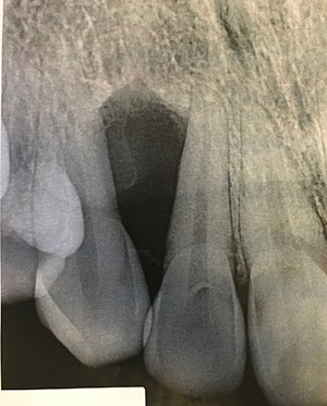
October 2019: Triangular radiolucent lesion between teeth #s 5 & 6
Can you make the correct diagnosis?
This is a 27-year-old female who had an incidental bony lesion between teeth #s 5 & 6. This lesion was asymptomatic and did not cause expansion in the area.
Sorry, you are incorrect!
The well-demarcated unilocular radiolucency, no expansion and very mild to no displacement of teeth should include odontogenic keratocyst (OKC) high on the differential diagnosis (DDX). The age of the patient is also consistent with OKC but as much the gender since OKCs tend to be slightly more common in males. The site however argues against OKC, since the majority occur in the posterior mandible and ramus area. The histology is also not consistent with OKC.
OKC is an aggressive odontogenic cyst and is known for its rapid growth and its tendency to invade the adjacent tissues, including perforating bone. It has a high recurrence rate and some (especially individuals under 15 years of age) are associated with basal cell nevus syndrome. OKCs typically occur in the age ranges of 20-29 and 40-59, but cases in patients ranging in age from 5 to over 90 years of age are reported. The distribution between sexes varies from equal distribution to a male-to-female ratio of 1.6:1, except in children. OKC predominantly affects Caucasian populations. OKC may occur in any part of the upper and lower jaw, with the majority of cases (almost 70%) occurring in the angle of the mandible and ramus. Radiographically, OKCs present predominantly as unilocular radiolucencies with well defined or sclerotic borders. OKCs of the maxilla are smaller in size when compared to those occurring in the mandible; larger OKCs tend to expand bone, but mildly—obvious clinical expansion should be viewed with suspicion for a neoplasm. OKCs have high recurrence rate, ranging from 13% to 60%. Complete surgical removal is the treatment of choice. Surgery combined with Carnoy’s solution or liquid nitrogen cauterization has been effective in reducing the recurrence rate. Most OKCs recur within the first three years while others may recur as late as after 26 years. Conservative surgical removal and long-term follow-up is the treatment of choice by most clinicians.
Sorry, you are incorrect!
The well-demarcated unilocular radiolucency and age of this patient are consistent odontogenic Myxoma (OM). The site and gender can be consistent with OM or argue against OM depending on who’s manuscript one is reading. Some investigators suggests that Oms are slightly more common in the maxilla and in females while others suggest posterior mandible and males being more common site and gender. The lack of expansion and displacement of teeth argue against OM. The histology is also not consistent with OM.
Odontogenic myxoma occurs in the jaw bones, usually in the tooth-bearing areas of the jaws. It is an uncommon, benign, but locally aggressive neoplasm. Nearly all cases so far have been described in the jaw bones. It is believed to be from the mesenchymal portion of a tooth germ, most likely of the dental papilla. It has the potential for extensive bony destruction and perforation into the surrounding soft tissue structures. They constitute around 17% of all odontogenic tumors. Almost 75% of odontogenic myxomas occur in patients around 23-30 years of age with a slight female predilection (1:1.5 male-to-female ratio). It rarely occurs in patients over 50 or under 10 years of age. It occurs almost equally in the maxilla and mandible with a slight predilection for the posterior mandible. A few cases are described in the ramus and condyle, which are non-tooth bearing areas. OM is a slow-growing, persistent and destructive but benign neoplasm. Most cases are expansile and can displace and resorb teeth. In the maxilla, they usually invade the maxillary sinuses and at times (though rarely) cross the midline to the opposing sinus. Radiographically, the majority present as expansile and multilocular, though some are unilocular with or without scalloped borders, and rare cases present with a diffuse and mottled appearance which can be mistaken for a malignant neoplasm. Grossly, this lesion is gelatinous in nature, making curettage alone difficult; the more fibrotic odontogenic myxomas (also known as odontogenic myxofibroma or fibromyxoma) have more body and are easier to curette. The treatment of choice is surgical excision ranging from segmental resection with clear bony margins of up to 1.5cm to prevent recurrence of the neoplasm. Curettage with and without cauterization is used for treatment, but is associated with a higher recurrence rate compared to the resected lesions.
Congratulations, you are correct!
The well-demarcated radiolucency that is triangular in shape anterior maxilla should place squamous odontogenic tumor (SOT) high on the DDX list. The age and gender can apply as well since SOT has no gender preference and can occur at a wide age range. The lack of pain is not typical of SOT since the majority of these neoplasms present with some pain. The histology is that of SOT.
SOT can be mistaken for acanthomatous ameloblastoma or intrabony squamous cell carcinoma. It is particularly important to differentiate between the clinical and radiographic findings of an ameloblastoma and intrabony squamous cell carcinoma from that of SOT. The latter is a non-aggressive benign neoplasm that can occur at any age (with a range of 11-67) with equal gender distribution. Both jaws are affected equally and it is usually associated with mild pain and tenderness. About 25% of cases are asymptomatic. Tooth mobility and multiple lesions have been described but is rare. Radiographically, it usually presents as a well-demarcated radiolucency, sometimes semicircular or triangular in shape. It is usually not corticated. These features are readily distinguishable, both clinically and radiographically, from a solid ameloblastoma as well as a malignant intrabony epithelial neoplasm. Simple curettage is adequate treatment for this lesion and has a good prognosis.
Sorry, you are incorrect!
The well-demarcated unilocular radiolucency, the site of maxilla anterior to the first molar and age of the patient are all consistent with the presentation of calcifying odontogenic cyst (COC). The lack of expansion is not unusual for the simple cystic COC but not the end of spectrum of ghost cell odontogenic tumor where it is usually expansile and can be aggressive. The histology is not consistent with COC.
The calcifying odontogenic cyst (COC) is a benign lesion of odontogenic origin. It has a spectrum of both clinical and histologic presentations that range from a simple cyst to an aggressive solid odontogenic epithelial neoplasm. The latter is also known as ghost cell odontogenic tumor (GCOT). The cystic counterpart (COC) constitutes around 1% of all odontogenic cysts.
COC tends to occur around the third decade of life, with an age range of 7-82 years. It occurs equally in the maxilla and mandible, usually anterior to the first permanent molar, though it has a predilection for occurrence in the maxilla in the younger age range. It occurs equally in males and females. GCOT, however, favors the mandible over the maxilla, usually anterior to the first permanent molar, but more commonly in males over fifty years of age. It occurs more commonly in men over 50 years of age. The behavior of GCOT is more persistent than that of its cystic counterpart. It tends to recur more often, and with multiple recurrences, it may transform into a malignant neoplasm. It is also important to mention that both COC and GCOT may occur in bone (more commonly) or in soft tissue (in the gingiva without a bony component, also known as peripheral COC or peripheral GCOT). The peripheral counterpart of both lesions is less aggressive than the intra-osseous type. Radiographically, COCs tend to be well defined radiolucencies with or without small flecks of radiopacities. They can be present at the apex, between teeth, or associated with impacted teeth. Clinically, both COC and GCOT can expand the jaws and extend into the surrounding soft tissues and the maxillary sinus. They can also resorb and displace teeth. Treatment ranges from thorough curettage to resection. The latter is suggested for the recurring and more aggressively behaving lesions.