Well demarcated mixed radiolucent/radiopaque lesion between teeth #s 22 & 23
Can you make the correct diagnosis?
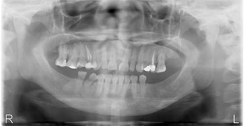
This is a 53-year-old female who presented with an asymptomatic swelling in the left anterior mandible between teeth #s 22 and 23.
Sorry, you are incorrect!
A well demarcated/corticated unilocular radiolucency is by definition the most common presentation for odontogenic cysts and therefore should be considered first on the differential diagnosis. The differential in this case would carry a periapical cyst, odontogenic keratocyst (OKC) and glandular odontogenic cyst (GOC). A periapical cyst would be unlikely since the involved teeth are vital. Odontogenic keratocyst can stay on the differential diagnosis but is usually not expansile and it more commonly occurs in the posterior mandible and ramus than anterior mandible. As to GOC, the clinical and radiographic presentation is consistent with it since it more commonly occurs in the anterior mandible and is usually expansile. The age of the patient is also consistent with GOC but not the gender. Therefore of three cysts considered, GOC would be the more likely one to present in this manner. The histology however was not consistent with periapical cyst, OKC or GOC.
Sorry, you are incorrect!
Benign cemento-osseous lesions should be considered on the differential diagnosis since they present looking radiolucent at the early stages of development. However, one can argue that in this case there is a hint of an early calcification given the cloudy appearance at the periphery of the unilocular lesion. Focal or periapical cemento-osseous dysplasias are usually unilocular and well demarcated. The age of this patient is not consistent with cemento-osseous dysplasia in general. The gender and the absence of clinical symptoms are consistent with it. The site and the more than one tooth involvement would be consistent with periapical cemento-osseous dysplasia (PCOD). The clinical and radiographic features of expansion and moving of the teeth apart are not consistent with PCOD. The histology is also not consistent with cemento-osseous dysplasia.
Sorry, you are incorrect!
The age of this patient is on the older age range for central ossifying fibromas (COF) while the gender and the clinical and radiographic presentation of expansion and moving of teeth apart is consistent with an early stage of COF. The histology in this case is not consistent with COF
Congratulations, you are correct!
A well-demarcated, expansile and moving teeth apart radiolucency in the anterior mandible should make one think of desmoplastic ameloblastoma and squamous odontogenic tumor. They both occur in the anterior jaws and are expansile and can move teeth apart. The age is consistent with both conditions. However, the histology was not consistent with either of the two neoplasms.
Another neoplasm to consider would be central odontogenic fibroma which can move teeth apart and is expansile and is usually radiolucent at the early stages of development. The site is unusual since they usually present in the anterior maxilla or posterior mandible. The age range is consistent with adult type of central odontogenic fibroma and the gender is consistent with it. The histology in this case was consistent with central odontogenic fibroma.