Unilocular radiolucency between teeth #s 28 & 29
Can you make the correct diagnosis?
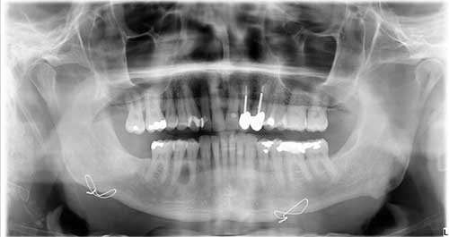
This is a 55-year-old white male who was referred to Dr. Kim for the evaluation of a slowly growing swelling in the right posterior mandible.
Sorry, you are incorrect!
A unilocular, well-demarcated/corticated radiolucency between teeth should bring to mind a tooth-related cyst; in this case, a variety of odontogenic cysts can fit this clinical presentation. The unilocular radiolucent lesion involves the entire length of the root, making the diagnosis of odontogenic keratocyst (OKC) more likely, but the site also triggers a differential diagnosis of OKC and a larger lateral periodontal cyst (LPC) or its variant botryoid odontogenic cyst (BOC). The latter is usually a multilocular radiolucency but can be unilocular.
OKC is high on the differential diagnosis because the posterior mandible is a common location for it. The well-demarcated/corticated radiolucent lesion is also a typical radiographic presentation for OKC. The age of the patient is on the older side of the age range, but OKC can occur at any age; this pathologist has noted an age range of 5 to 92. An additional factor supporting a clinical diagnosis of OKC is the gender of the patient, since OKC is slightly more common in males. The histology, however, is not consistent with OKC.
Congratulations, you are correct!
Lateral periodontal cyst (LPC) and its variant botryoid odontogenic cyst (BOC) are also high on the differential diagnosis. These two cysts tend to occur between cuspids and bicuspids and are more common in males over the age of 30. Therefore, the age, gender, and site is consistent with both cysts. Radiographically, LPC usually presents as a small, corticated and unilocular radiolucency between teeth at the middle to superior roots, rather than involving the entire length of the root as is the case in this patient. However, rare LPCs can involve the entire length as well. BOC, a variant of LPC, is usually a multilocular radiolucency but can be unilocular. BOCs usually involve the full length of the root. They are more aggressive in behavior than LPC. The histology in this case is consistent with LPC with a note that BOC is on the differential diagnosis given the size of the lesion. Follow up visits were recommended.
Sorry, you are incorrect!
The posterior mandible is a common location for a variety of benign odontogenic neoplasms, especially those in the ameloblastoma family. Solid ameloblastomas, however, tend to be multilocular and tend to push posteriorly into the ramus. They are also obviously expansile and aggressively pushing teeth apart; none of that is noted in this patient’s clinical and radiographic changes.
The unicystic variant of ameloblastoma usually presents as a unilocular, well-demarcated to corticated radiolucency and therefore can be considered on the differential diagnosis. Support for the diagnosis of unicystic ameloblastoma is not strong because unicystic ameloblastomas typically occur in patients around 10-20 years of age, presenting 90% of the time in the posterior mandible associated with impacted third molars and radiographically looking like a dentigerous cyst. The other 10% of cases can present between teeth as unilocular radiolucencies, as in this case, but the age of this patient is too old for that condition to be likely. The histology is not consistent with solid or unicystic ameloblastoma.
Another odontogenic neoplasm to consider, but not high up on the list, is squamous odontogenic tumor (SOT). This benign neoplasm/hamartoma of epithelial odontogenic origin can occur anywhere in the jaws, including between the premolar and molars. Radiographically, SOTs are usually semicircular or triangular with a corticated border. This lesion is radiographically fully circular, making it less likely to be an SOT. SOT can occur at any age, particularly in adults over 40 years of age, with no gender predilection. This patient’s lack of clinical symptoms such as pain or tooth mobility argues against a diagnosis of SOT. The histology is not consistent with SOT.
Sorry, you are incorrect!
CGCG should be considered on the differential diagnosis because of the site anterior to the first molar. However, the age and gender of the patient are not consistent with CGCG. About 60% of CGCG cases occur in patients under 30 years of age. The radiographic features of this case are also not typical of CGCG; it is more commonly a multilocular radiolucency rather than a corticated unilocular radiolucency, though the latter can be the presentation on rare occasions. Also, radiographically, the margins of CGCG tend to be less defined. The histology in this case is also not consistent with CGCG.