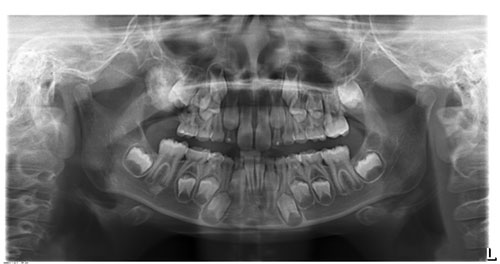
Predominantly radiopaque lesion associated with unerupted tooth #3
Can you make the correct diagnosis?
This is a 7 1/2 year old male who presented with tooth #3 unerupted. There was no palpable expansion of the right posterior maxilla.
Disclaimer: It is important to state that the differential diagnosis (DDX) of this case should focus on tooth-related conditions. The rationale for this approach is that this lesion is associated with the unerupted tooth #3 which makes non-tooth related pathology unlikely. The DDX will be limited to those conditions (cysts and neoplasms) that are of tooth origin and are known to produce odontogenic hard tissue.
Sorry, you are incorrect!
Radiographic findings of a well-demarcated and large radiopaque mass associated with an unerupted tooth should lead one to begin the DDX with the most common odontogenic mixed RL/RO lesion, odontoma. In this case, it would be a complex odontoma because it is in the form of a large opaque mass associated with an unerupted tooth #3 in the posterior jaw. The patient’s age is within the typical age range of this condition, albeit on the younger side. The fact that the mass is associated with an unerupted/impacted molar tooth is also consistent with this condition, as are the radiographic findings. The location in the posterior jaw is consistent with this condition, but complex odontomas tend to be more common in the posterior mandible associated with third molars. The lack of expansion, as in this case, is reported in some odontomas.
Complex odontomas are usually associated with unerupted teeth in the posterior jaws. They are more often associated with third molars than first or second molars. They can occur at any age but are more common in the first two decades of life. The histology in this case is partially that of an odontoma.
Congratulations, you are correct!
The clinical presentation of a delayed eruption of tooth #3 and the association of this tooth with a well-demarcated RL/RO lesion should lead one to consider ameloblastic fibro-odontoma (AFO) on the DDX. AFOs tend to occur in children around 10 years of age, close to the age of this patient. These lesions are slow growing and can reach large sizes, as is the case in this patient. The site in the posterior maxilla and the patient’s gender are also consistent with AFO since they have equal gender and equal jaw distribution. Some reports indicate that AFO may have a slight predilection for occurrence in the posterior maxilla, as is the case in this patient. The radiographic findings of a well-circumscribed and expansile mixed RL/RO mass are also consistent with AFO. The histology in this case is that of AFO.
Sorry, you are incorrect!
Calcifying odontogenic cysts (COC) generally occur in patients around 30 years of age with equal gender distribution and equal rates of occurrence in each jaw. This clinical profile changes when COCs are associated with odontomas (COC+O). Between 22% and 47% of COCs are associated with odontomas, including complex odontomas. The mean age of occurrence for COC+O is 16, and it is more common in females (F:M ratio 2:1) and slightly more common in the maxilla. The site in this case is consistent with COC+O, but not the patient’s age or gender. Nor is the association with impacted teeth common; fewer than 30% of COC+O cases are associated with impacted teeth. Radiographically, COC+Os are well-circumscribed RL with RO flecks or masses depending on whether it is COC alone or COC+O. The histology, however, is not consistent with COC. Histology shows an odontoma but no evidence of COC.
Sorry, you are incorrect!
Pindborg tumor was first described in 1956 and constitutes less than 1% of odontogenic tumors. It occurs equally across genders. However, the age of this patient is too young for this condition since Pindborg tumors rarely occur in patients under 10 years of age. The typical age range for Pindborg tumor is 30-50 years. The site of occurrence is not consistent with Pindborg tumor since close to 75% of Pindborg tumors occur in the posterior mandible. Slightly more than 50% of CEOTs are associated with impacted teeth. In this case, it is too early to describe the status of tooth #3 as impacted; it may just be delayed eruption due to the mechanical pressure imposed by the large lesion. Tooth #3 was saved (Figure 3) to allow it to erupt. Radiographically, CEOT presents in several patterns, including a well-circumscribed unilocular radiolucency with radiopaque masses as is the case in this patient. It can also present as a multilocular “honeycomb” radiolucency with flecks of radiopaque material, a so-called “driven snow” pattern. The histology of this case, however, is not consistent with Pindborg tumor.