All cases are discussed by: Dr. Dolphine Oda, UW-Oral Pathology Biopsy Service
Diffuse gingival redness/erosions and sloughing
Contributed by: Dr. Terry LaBell
LaBell Periodontics & Implants, Woodinville, WA
Case Summary and Diagnostic Information
This is a 47-year-old male who presented with diffuse gingival redness since late October 2017; initially started in the lower left canine area and progressively spread throughout the dentition.
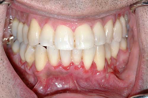
This is a 47-year-old male who presented with diffuse gingival redness since late October 2017; initially started in the lower left canine area and progressively spread throughout the dentition (Figure 1). Patient reported the lesions were painful when eating or brushing. Upon presentation to the periodontist, there was an intense erythema on the facial of teeth #12-13, 19-25 and 28-29. The most intense redness was on #20-25 (Figure 2). The tissue bled easily with any contact and exhibited a positive Nikolsky sign.
Figure 1 This is a clinical photograph taken of the patient when he presented to the periodontist. Note the diffuse red/eroded left and anterior mandibular gingiva and sloughing epithelium.
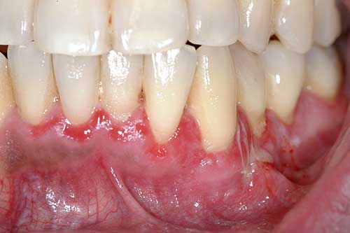
Figure 2 This is a closer look at the left and anterior mandibular gingiva demonstrating the diffuse red and eroded gingiva with clearly sloughing epithelium.
The patient’s past medical history is significant for benign atrial fibrillation and non-alcoholic fatty liver disease. He reports rare alcohol consumption and no use of any tobacco products or recreational drugs. Current medications include propranolol and fish oil.
In late October 2017, this patient complained about red painful gingival lesions bleeding when brushing. Initially, the lesions were confined to gingiva of tooth #22 which over a six month period, it progressively spread on other parts of the mandibular gingiva and also involved the maxillary gingiva.
Under local anesthesia, two incisional biopsies were taken one placed in formalin and the second in Michel’s solution for immunofluorescent staining. At biopsy, the area bled profusely and appropriate measures were taken to stop the bleeding.
Histologic examination reveals one piece of soft tissue composed of surface epithelium splitting above the basal cell layer (Figure 3-5). The spinous layer shows evidence of acantholysis (Figure 5) and the basal cell layer is intact and part of the connective tissue (Figure 4). The latter is infiltrated by a mixed inflammatory population. Direct immunofluorescent staining shows positive intercellular staining of the spinous layer cells with antibody to IgG and C3 (not shown).
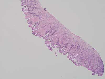
Figure 3 This is a low power (40X) photograph of an H & E stained section, demonstrating mostly detached epithelium above the basal cell layer. The basal cells are intact and clearly anchored on the basement membrane. The detached epithelium shows evidence of acantholysis. The connective tissue is infiltrated by mixed and chronic inflammatory cells.
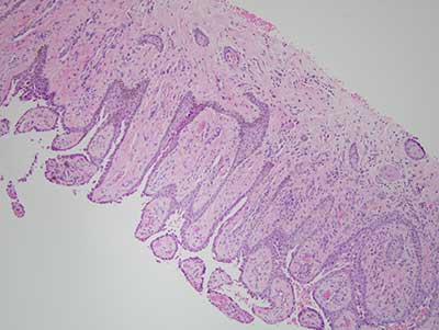
Figure 4 This is a higher power (100X) photograph of an H & E stained section clearly demonstrating the epithelial detachment above the basal cell layer.
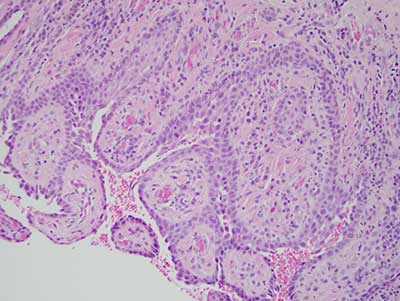
Figure 5 This is high power (200X) photograph of an H & E stained section, focusing on the split of the surface epithelium, the breakdown of the spinous layer and the acantholytic cells
After you have finished reviewing the available diagnostic information