Diffuse gingival redness/erosions and sloughing
Can you make the correct diagnosis?
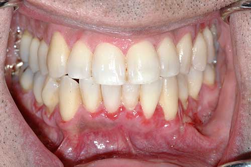
This is a 47-year-old male who presented with diffuse gingival redness since late October 2017; initially started in the lower left canine area and progressively spread throughout the dentition.
Differential Diagnosis
When erosions, ulcerations, epithelial sloughing are confined to the gingiva only and not anywhere else in the mouth, skin or other mucosae, the applied and more likely differential diagnosis shrink to a few conditions including:
Sorry, you are incorrect!
This is a chronic immune mediated disease predominantly affecting the oral cavity especially the gingiva. Three types are described, the chronic oral type known as oral mucous membrane pemphigoid, the eye subtype causing scarring as cicatricial pemphigoid and the skin (commonly head and neck area) with bullous formation as the bullous pemphigoid.
This case shows over a six month period progressive red gingival patches with loss of stippling and sloughing epithelium in an adult patients all of which should make one start with immune mediated diseases such as mucous membrane pemphigoid (MMP). The age, the site and chronic and progressive change are all consistent with MMP. The gender however is not since oral MMP tends to be twice as common in females as males. MMP tends to occur in individuals over 40 years of age, but is described in children but rarely. Up to 94% of MMP cases occur on the gingiva than anywhere else. If the gingival lesions are untreated, they will slowly involve other areas in the mouth, eyes, larynx, esophagus and others. Positive Nikolsky sign and sloughing of epithelium in response to mechanical pressure such as from brushing is consistent with the clinical behavior of MMP. Both the H & E histology and the direct immunofluorescence staining are negative for MMP.
Sorry, you are incorrect!
Typically, oral lichen planus (LP) and its family of diseases represent chronic conditions with a variety of etiologies including immune mediated. In the oral cavity, they tend to present in a generalized and symmetrical manner involving the bilateral buccal mucosa and vestibule, dorsal and lateral tongue and gingiva which argues against this case being lichen planus or lichenoid mucositis. However, about 10% of LP cases occur only/mostly on the gingiva. That presentation is usually lichenoid reaction to something a medication or to restorative dental material.
In this case, gingiva is the only site affected which makes lichenoid mucositis more likely than the conventional erosive lichen planus. Lichenoid mucositis are more common in elderly patients, more in females. The age and gender in this case argue against lichenoid mucositis, so does the histology and DIF staining pattern.
Congratulations, you are correct!
Pemphigus vulgaris is a rare family of serious acantholytic/blistering diseases. This is a progressive and chronic immune mediated disease where the immune system recognizes certain proteins in the desmosomes to be antigenic and destroys them causing breakdown of the spinous layer cells (acantholysis) forming blisters within the epithelium ultimately leading to break down of the epithelium and causing ulceration. This disease affects the skin and mucosa including oral (about 50% of cases), eyes, upper respiratory, genitals and other areas. Pemphigus vulgaris occurs more commonly in females 40-60 years of age. In the oral cavity, it is more common on the lips, and soft palate area. The gender of this patient is not consistent with PV and neither is the site. The clinical presentation of slow progression of the disease and the age are consistent with PV and so is the histology and the direct immunofluorescence staining.