Large & Well-Demarcated Radiolucency Left Posterior Mandible
Can you make the correct diagnosis?
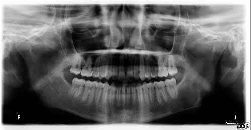
This is a 15-year-old white female with a large radiolucency in the left posterior mandible and ascending ramus.
Sorry! you are incorrect
Given the radiographic findings of a unilocular and well-defined radiolucency, a cyst has to be on the differential diagnosis. OKC is considered high up on the differential diagnosis given the lack of expansion but the histology is not supportive of OKC.
The odontogenic keratocyst (OKC) is an aggressive odontogenic cyst, known for its rapid growth and its tendency to invade the adjacent tissues, including bone. It has a high recurrence rate and is associated with basal cell nevus syndrome. It affects patients in the age ranges of 20-29 and 40-59, but cases in patients ranging in age from 5 to 80 years have been reported. The distribution between sexes varies from equal distribution to a male-to-female ratio of 1.6:1, except in children. Odontogenic keratocysts may occur in any part of the upper and lower jaw, with the majority (almost 70%) occurring in the mandible. They occur most commonly in the angle of the mandible and ramus. Anterior maxilla is a rare location for OKC but has been described.
Radiographically, OKCs present predominantly as unilocular radiolucencies with well-defined or sclerotic borders; they may also present as multilocular radiolucencies, but rarely. OKCs commonly present as a unilocular radiolucency with scalloped borders. Teeth associated with OKC are vital. OKCs grow to sizes larger than any other odontogenic cysts. They usually penetrate the bone rather than expand it and grow in an anterior to posterior direction. Despite this aggressive growth, they often remain asymptomatic, thus growing to large sizes and hollowing the bone. Treatment of choice is surgery with cauterization, especially with Carnoy’s solution.
Sorry! you are incorrect
A well-demarcated radiolucency in the vicinity of the inferior alveolar nerve and canal should make one think of a benign neoplasm of peripheral nerve sheath origin such as schwannoma or neurofibroma. The age of the patient, the location and the radiolucency are all supportive of this diagnosis. However, the panoramic radiograph alone is not adequate imaging in determining origin from the alveolar canal. The lack of expansion is also not consistent with this neoplasm. The histology is not supportive of this neoplasm.
Schwannoma, also known as neurilemmoma is a benign, encapsulated neoplasm of Schwann cell origin. It is mostly a soft tissue neoplasm that is rare in the mouth. The soft tissue schwannomas are by far more common than the intra-osseous schwannomas. The soft tissue lesions tend to occurs in young and middle aged patients with equal sex distribution. Tongue is the most common location but it can also occur in the lips, buccal mucosa and floor of mouth. The intra-osseous schwannomas are rare but the head and neck area is the most common location for these neoplasms constituting up to 48% of the intra-osseous schwannomas. They are more commonly described in females (2:1) average age of 29. A significant number of these cases occur in children under 12 years of age. They are slow-growing and can expand the jaw. They are by far more common in the posterior mandible and ramus and rarely occur in the maxilla. They can be associated with pain and paresthesia. Histologically both types, the soft and intra-osseous have the same morphology characterized by two patterns: Antoni A, the cross section of which gives rise to what is called “Verocay bodies,” and Antoni B, which is loose and resembles neurofibroma. The treatment of choice is conservative surgical excision.
Congratulations! You are correct
The well-demarcated non-expansile, unilocular radiolucency with scalloped border in the mandible of a young person should make one think traumatic bone cavity (TBC) and this was the diagnosis. Also, note the demarcation of the radiolucency, it is corticated or clearly defined in some areas. This would be more consistent with TBC. However, the very posterior presence and extension into the ramus of the mandible is not typical of TBC.
The traumatic bone cyst is best called a traumatic bone cavity since this condition does not represent a true cyst. Traumatic bone cavity (TBC) is not unique to the jawbones; it is also described in the long bones and is known as a simple solitary bone cyst occurring mostly in the humerus or femur, close to the epiphyseal plate. The long bone simple cyst is similar to the jaw traumatic bone cavity radiographically and occurs in younger age range. Trauma has been suggested as the etiology along with other non-substantiated theories such as cystic degeneration of a preexisting tumor or of the fatty marrow in the area.
Some reports suggest that it is more common in males while others report equal distribution between males and females. The long bone counterpart is more common in males by a ratio of 2.5:1. Most reports agree that the average age of occurrence is below 20 years of age. These lesions can occur, but are uncommon, over the age of 30. Kaugars reported a higher number of TBC cases in African-American females compared to the literature. The latter patients were over the age of 30. This may suggest an association with florid cemento-osseous dysplasia. The mandible is the most commonly affected area, where over 95% of cases occur, especially in the posterior premolar-molar area. TBCs are also known to cross the midline anteriorly. In one study, 27% of cases were anterior to the canine and some crossed the midline. They are usually unilocular and radiolucent, typically above the alveolar canal, and in many cases have a scalloped superior border squeezing between the roots of teeth. The latter are vital and are frequently found hanging within the empty cavity. About 25% of the lesions occur in the anterior mandible apical to the canine tooth and are usually round and unilocular; they can therefore be mistaken for periapical lesions, leading to an unnecessary endodontic treatment. Therefore, it is important to test the vitality of the teeth and carefully examine the radiographs for changes consistent with a periapical granuloma or cyst. Though expansion is not characteristic of TBC, it is described in about 26% of cases. TBCs are otherwise asymptomatic. The margins of these lesions range from very well defined to corticated to punched-out radiolucency. Pathologic fractures associated with traumatic bone cavity have been described in the jaws, but are rare. They are, however, more common in association with TBCs of the long bones.
Clinically, surgeons report an empty cavity at entrance in about two thirds of cases and cavities filled with straw-colored fluid in about one third of cases. Blood clots are also present occasionally. The bone cavity is scraped to generate bleeding, which is considered the treatment of choice for this condition. Other methods of treatment have been tried, such as packing the curetted cavity with autogenous blood, autogenous bone and hydroxyapatite. Various other reports demonstrate healing of TBC after injection of autogenous blood, after aspiration and after endodontic treatment. These lesions may spontaneously heal, but rarely. Biopsy material consists of fragments of viable bone and loose connective tissue. Osteoclast-like giant cells have also been described in a few cases. Exploration surgery usually leads to healing. Recurrence is rare.