Large Radiolucency with Scalloped Border Left Posterior Mandible
Dolphine Oda, BDS, MSc
doda@u.washington.edu
Dr. Sasi Narra
Oral & Maxillofacial Surgery, Issaquah, WA
Case Summary and Diagnostic Information
This is a 15-year-old white female with a large radiolucency in the left posterior mandible and ascending ramus.
Diagnostic Information Available
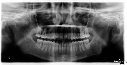
This is a 15-year-old white female with a large radiolucency in the left posterior mandible and ascending ramus (Figure 1). It was an incidental finding. The lesion is of unknown duration and is asymptomatic. The radiolucency is not expansile. The past medical history is unremarkable.
Figure 1. This is a panoramic radiograph taken at first clinical presentation. Note the large, well-defined unilocular radiolucency with scalloped border in the posterior mandible extending into the ascending ramus.
The patient’s past medical history is negative for any significant diseases or risk factors.
The lesion is an incidental finding and is of unknown duration. There was no pain or paresthesia or any other symptoms associated with this radiolucency. The radiolucency was not expansile.
Treatment
Under local anesthesia, a full-thickness flap around the area was raised and the area was scrapped. There was some straw-colored fluid but no solid tissue. The area was sutured and the specimen submitted for microscopic evaluation.
Excisional Biopsy
Histologic examination reveals multiple pieces of hard and soft tissue composed of bone, fibrous connective tissue with small blood vessels, extravisated erythrocytes and small fragments of peripheral nerve bundles (Figures 2 & 3). Bone comprises the bulk of the specimen and is viable. Inflammatory cells are absent.
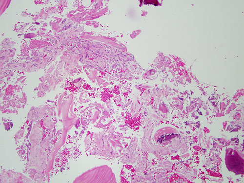
Figure 2. Low power (x100) H & E histology illustrates fragments of bone interspersed with fibrous connective tissue containing small blood vessels, erythrocytes, and small bundles of peripheral nerve fibers.
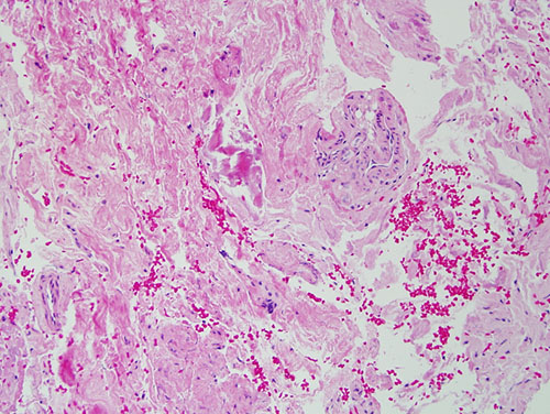
Figure 3. Higher power (x200) H & E histology illustrates higher power of the fibrous connective tissue with small blood vessels and extravisated erythrocytes.
After you have finished reviewing the available diagnostic information