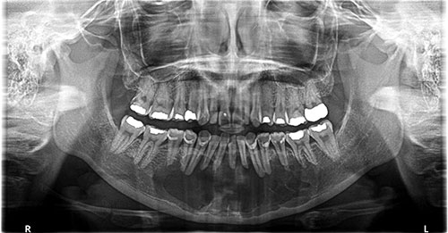
Large multilocular, expansile radiolucency, anterior mandible
Can you make the correct diagnosis?
This is a 30-year-old female presented to her dentist, as a new patient, with an expansile swelling in the anterior mandible.
Congratulations, you are correct!
The site of anterior mandible and the radiographic findings of a well-demarcated; expansile and multilocular radiolucency are consistent with the clinical and radiographic presentation of GOC. The age on the other hand is on the younger age range and the gender is not consistent since GOC is slightly more common in males. The histology is that of GOC.
Glandular odontogenic cyst (GOC) is a developmental cyst of tooth origin characterized by unusual lining epithelium and occasional aggressive behavior. The uniqueness of this cyst lies in its histology. The lining epithelium is stratified squamous in type, but covered by cuboidal or columnar cells (sometimes ciliated) interspersed with microcytic spaces simulating salivary gland ducts, giving rise to the name “glandular.” It is a rare cyst and, though mostly inert, it can sometimes be aggressive in behavior. Because it occurs in association with teeth, it is believed to be of tooth origin. Therefore, the name “glandular” is misleading. Glandular odontogenic cyst is more common in adults of an average age of 49, with a slight male predominance. It has, however, been reported in all age ranges, including teenagers. It occurs three times more commonly in the mandible than in the maxilla, especially in the anterior mandible. Radiographically, they tend to present as unilocular and, less commonly, as multilocular radiolucencies. Multilocular GOCs tend to recur more than unilocular ones. These lesions grow to large sizes in the majority of cases and can perforate bone in a manner similar to the behavior of odontogenic keratocyst, a known aggressive cyst of tooth origin. Like OKCs, GOCs can be aggressive in bone perforation and recurrence rate. The recurrence rate ranges between 21-55%. Enucleation and curettage alone carry a high recurrence rate of 25%. Marsupialization has been successfully used.
Sorry, you are incorrect!
The gender, age, site, the crossing of the anterior mandibular midline and the radiographic findings of well-demarcated, expansile and multilocular radiolucency are all features consistent with those of CGCG. The histology, however, is not consistent with CGCG.
Central giant cell granuloma is a non-neoplastic condition that can occasionally behave in a very aggressive and expansile manner, destroying bone and displacing teeth. Over 60% of CGCG cases occur in patients younger than 30 years of age, with twice as many occurrences in females as in males. CGCG is classified into aggressive and non-aggressive types; the aggressive type tends to occur in younger patients and is known to cause disfiguration, especially after surgery. Over 70% of cases occur in the mandible anterior to the first molar tooth.
Sorry, you are incorrect!
The well-demarcated multilocular and expansile radiolucency is “typical” radiographic presentation of solid ameloblastoma. The patient’s age fits well into the mean age of this condition. Focal bone perforation is also consistent with the clinical behavior of solid ameloblastomas. Solid ameloblastoma occurs equally in males and females. The site however is not consistent with this condition. The histology is also not consistent with solid ameloblastoma.
Solid ameloblastoma is one of the most common benign neoplasms of odontogenic origin. It accounts for 11% of all odontogenic neoplasms. It is a slow-growing, persistent, and locally aggressive neoplasm of odontogenic epithelial origin. It affects a wide age range but is mostly a disease of adults with an average age of 33 and equal sex distribution. Reports from Africa and India show a male predilection; it also has a predilection for occurrence in black patients.
About 85% of ameloblastomas occur in the posterior mandible; most of these occur in the molar-ramus area, and some occur in the anterior mandible but sporadically. Solid ameloblastoma is characteristically expansile, radiolucent and multilocular in nature. Solid ameloblastoma, if not treated, can reach very large sizes, causing facial disfigurement. It can loosen, displace and resorb adjacent teeth and perforate bone. With the exception of jaw expansion, it is usually asymptomatic unless infected, in which case it can be mildly painful. Paresthesia and anesthesia are very rare, unless the lesion is very large in size. It has a high tendency for recurrence if not treated appropriately with clean margins.
Sorry, you are incorrect!
Generally, the site and the multilocular and expansile radiolucency argue against OKC since this cyst more commonly occurs in the posterior mandible and ramus. The age of this patient is within the age range. As to the multilocular radiographic presentation, around 10-12% of OKCs can be multilocular and those are usually large. The larger and multilocular types of OKC tend to expand the jaw bone and move teeth apart. The areas of bone perforation are also consistent with the behavior of OKC. The histology is not consistent with OKC.
Odontogenic keratocyst is an aggressive cyst known for its rapid growth and its tendency to invade the adjacent tissues and perforate bone. It has a high recurrence rate and is associated with bifid rib basal cell nevus syndrome. The majority of patients are in the age ranges of 10-40 years of age with a wide age range of 5-92. The distribution between sexes varies from equal distribution to a male-to-female ratio of 1.6:1, except in children. OKC predominantly affects Caucasian populations chiefly of Northern European descent.
The majority (almost 70%) of OKCs occur in the angle of the mandible and ramus. Radiographically, OKCs present predominantly as unilocular radiolucencies with well-defined, sclerotic or scalloped borders. They may also present as multilocular radiolucencies. The larger OKCs tend to expand bone, but mildly—obvious clinical expansion should be viewed with suspicion for a neoplasm. OKCs can also present as small and oval radiolucencies between teeth simulating a lateral periodontal cyst, in an area of an extracted tooth simulating a residual cyst, at the apex of a vital tooth mistaken for a periapical cyst, or in the anterior maxilla between the central incisors simulating an incisive canal cyst. OKCs grow to sizes larger than any other odontogenic cysts. They usually penetrate the bone rather than expand it and grow in an anterior to posterior direction. Despite this aggressive growth, they often remain asymptomatic, thus growing to large sizes and hollowing the bone. They have a recurrence rate of 12% to 60%. Most cysts recur within the first three years while others may recur as late as after 26 years (this reviewer’s experience).