Expansible multilocular radiolucency: Left posterior mandible
Contributed by Drs. Srinivasa Chandra & Darrell Tew
Oral & Maxillofacial Surgery, Harborview Medical Center & Yakima, WA
Case Summary and Diagnostic Information
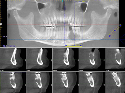
This is a 50-year-old Hispanic male who presented with a lesion of less than a year’s duration in the posterior mandible.
This is a 50-year-old Hispanic male who presented with a lesion of less than a year’s duration in the posterior mandible. His symptoms included pain, 3+ tooth mobility, 15-pound weight loss and swelling of the gingiva and jaw. The patient also complained of anesthesia of the left chin and lower lip. The patient had been aware of the symptoms with facial swelling of the left side for only a few months (Figure 2). Dr. Tew extracted the mobile teeth and biopsied the area. The initial cone beam CT scan findings (Figure 1) notably show a well-demarcated multilocular and expansile radiolucency perforating bone and pushing teeth apart.

Figure 1 This is an image of a cone beam CT scan which shows the complete bicortical destruction of the entire height of the mandible between teeth #s 19 & 22 with a multilocular and expansile presentation.
The patient’s past medical history was initially reported as “none known”; his last visit to a physician had been 15 years prior. On preoperative workup, he was found to have untreated hypertension and type 2 diabetes. He reported no surgical history. Family history is negative. He was a tobacco smoker of unspecified duration.
The clinical presentation was that of a left sided fullness (Figure 2). There was anesthesia of the left chin and lower lip. Intraoral clinical examination revealed buccal/lingual expansion of the posterior mandible. The bony enlargement was in area between teeth #s 19 to 22 with gross tooth mobility. Radiographic examination revealed bony expansion, with a lytic lesion of the left mandibular body and displacement of adjacent teeth roots (Figure 1).

Figure 1

Figure 2 This photograph is taken at first clinical presentation. Note the fullness of the left sided lower face.
The treatment consisted of enbloc resection and reconstruction with a composite osteocutaneous fibular free flap. Under general anesthesia and by a trans-cervical approach, the mandibular resection was performed after earlier intermaxillary fixation. A reconstructive titanium bar was placed and fixated. A right fibular free flap with a fascial skin paddle with the vascular pedicle was raised and osteotomy of the fibula was performed to fit into the 6.5 cm mandibular defect. Figure 3 shows the results 12 months after surgery, demonstrating mandibular osteo-integration of the fibula with the mandible held by a titanium reconstruction plate. Figures 4a & b show the intraoral mucosal healing of the skin paddle after cutaneous debridement and dental rehabilitation.
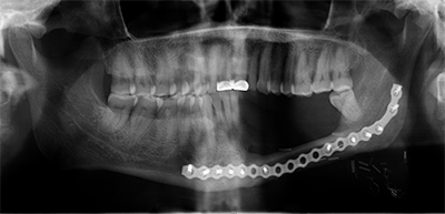
Figure 3 This photograph shows a 12-month postoperative panoramic radiograph with good osteo-integration of the fibula into the native mandible being supported by a titanium plate. The occlusion is intact and well preserved.
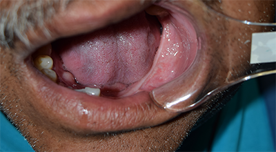
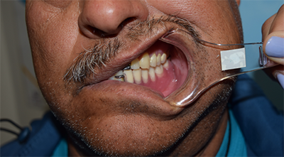
Figure 4a & b These photographs show the intraoral mucosal healing of the skin paddle after cutaneous debridement and dental alveolar ridge with a partial denture completing his dental rehabilitation.
Histological examination revealed multiple pieces of soft tissue made up of an odontogenic neoplasm of epithelial origin (Figures 5 a & b). This neoplasm is composed of many epithelial islands arranged in a spectrum of islands mostly containing squamous epithelium in the center to those with stellate reticulum in the center to very small nets and cords of neoplastic epithelial cells with no columnar border (Figure 5a). Most of the larger epithelial islands were lined by one layer of palisaded columnar cells some with reversed polarization (Figure 5b).
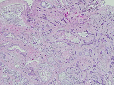
Figure 5a Low power (x40) H & E stained section revealing a neoplasm made up of epithelial islands, cords and nests suspended and surrounded by mature connective tissue. The center of the epithelial islands contains squamous, stellate or cuboidal epithelial cells.
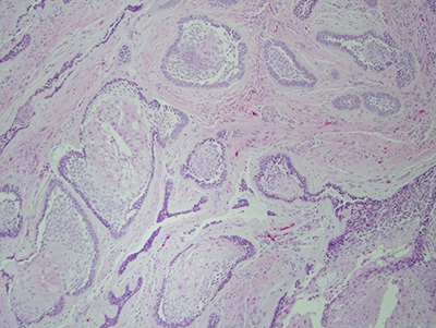
Figure 5b Higher power (x100) H & E stained section revealing the neoplastic epithelial islands surrounded by columnar cells, some with revered polarization.
After you have finished reviewing the available diagnostic information