Large exophytic swelling, right retromolar pad area and gingiva of tooth #31
Can you make the correct diagnosis?
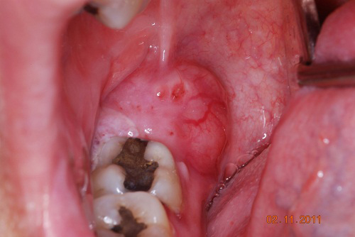
This is a 61-year-old white female who had a slowly and progressively enlarging gingival swelling over the last six months.
Sorry! you are incorrect
Granted, this is not a truly gingival swelling; it is mostly a retromolar pad swelling. It is, however, a swelling with a sessile base and lobular, exophytic, pinkish-red color suggestive of a pyogenic granuloma. The large size is unusual but could still reflect a PG. The histology is not supportive of PG.
Pyogenic granuloma constitutes 85% of all reactive gingival swellings but can occur in areas other than the gingiva. It represents a profuse mass of vascular granulation tissue. It can be induced by trauma and local irritants such as excessive plaque, sharp fillings and dental calculus; it sometimes forms in an extraction socket in response to an irritant left in the socket. It can occur anywhere in the oral cavity and skin, especially the tongue, lips, fingers and nail beds. In the mouth, it occurs most commonly in the gingiva, especially the maxillary buccal and interproximal gingiva. Occasionally, it may surround the tooth. It is usually highly vascular, fast-growing, exophytic, lobular, sessile, and ulcerated or covered by pseudomembrane. The color changes from red to pink when it starts to heal. It occurs at any age and sex with a slight predilection for young females; it affects 1% of pregnant females. Pyogenic granuloma is usually painless except during eating, when bleeding and pain is described. Histologically, it presents as a mass of loose and vascular granulation tissue, usually with ulcerated or eroded surface epithelium and many inflammatory cells. A range of treatment modalities are available, including excision with removal of the local irritant, laser surgery, or intralesional injection with absolute alcohol, steroids or botulinum toxin. Scaling and polishing prior to surgical removal helps shrink the lesion. The prognosis is good, although recurrence is possible, especially during pregnancy.
Sorry! you are incorrect
Although squamous cell carcinomas (SCCs) of the oral cavity rarely present in an exophytic and lobular manner, they do occasionally present in that form, especially those that occur on the gingiva. Again, this lesion is not truly of gingival origin but is in the proximity of tooth #31. The six-month duration, the lack of etiology and the red and white color suggests that SCC should be included on the differential diagnosis. The histology, however, is not supportive of SCC.
Squamous cell carcinoma of the gingiva is uncommon, especially in a non-smoker and social alcohol user. Oral squamous cell carcinoma of the mouth is a highly aggressive neoplasm that currently ranks as the fifth most common malignant neoplasm worldwide and accounts for an estimated 90% of oral malignancies. Oral SCC occurs predominantly in males over the age of 40 years, with an observed male-to-female ratio of 2:1 generally and 1.4:1 in the USA. Excluding the outer lip, the most common sites (in decreasing order) are the ventral and lateral surfaces of the tongue (25-50%), floor of mouth (15%), gingiva (12%) and palate (9%). The buccal mucosa and retromolar pad areas (3%) have a relatively low incidence of occurrence unless the patient is a chronic smokeless tobacco user. Oral SCC varies in presentation from deceptively innocent-looking to obviously malignant. It may present as a non-healing ulcer, or as red, white or mixed red-and-white lesions. Characteristic signs of oral SCC are non-healing ulcer, ulcer with rolled borders, fungation, fixation and induration. Rarely, oral SCC may present as unexplained asymptomatic lateral neck lymphadenopathy. Oral SCC is most commonly associated with chemically induced mutagenesis, specifically tobacco and alcohol use.
Tobacco use is described in over 75% of oral SCC patients. Tobacco and alcohol have been shown to act synergistically in the development of oral SCC. Human papilloma virus (HPV) has also been found to have a high prevalence in oral cancer, especially in younger patients with no history of tobacco use but more so in the posterior mouth, tonsil and oropharyngeal area. Other factors include poor oral hygiene, syphilis, chronic candidiasis, iron and dietary deficiencies, herpes simplex and various other immunologic factors, and lichen planus—especially persistent erosive lichen planus.
Determination of the prognosis of oral SCC is based on its clinical stage and histological classification. Although oral SCC is a diagnosis made by histology, surgeons tend to depend exclusively on the TNM classification system for clinical staging and treatment decisions. Prognosis is dependent on the TNM staging system; the most important prognostic sign is the presence or absence of metastases at the time of diagnosis. The prognosis thus improves when the lesion is detected early. Oral SCC patients die mainly of infection due to lowered resistance or of hemorrhage if the tumor erodes through one of the main blood vessels.
Sorry! you are incorrect
The most common location in the mouth in which metastatic cancer manifests is the posterior mandible, followed by the gingiva. Granted, cancer metastasis to the mouth is exceedingly rare and given that this patient has no history of primary cancer, it would be highly unlikely that this lesion represents a metastatic cancer. However, the six-month duration of a persistent disease is not conducive to definitively ruling out a metastatic disease. The histology is not supportive of a metastatic malignancy.
Cancer metastasis to the oral cavity is neither specific nor common. Although it constitutes fewer than 1% of all oral malignant neoplasms, it may have a devastating result to the patient mainly because metastasis to other sites has already developed or is inevitable. Theoretically, any malignant neoplasm can metastasize to the oral cavity, but in actuality few do and out of the ones that do, the majority are carcinomas rather than sarcomas. The most common malignant neoplasms that metastasize to the mouth are from the breast, lung, kidney and prostate. Malignant neoplasms from the thyroid, pancreas, colon, and liver have also been described. Breast cancer is the most common neoplasm to metastasize to the oral cavity regardless of gender. However, lung and prostate cancers are the most common neoplasms to metastasize to the oral cavity in men. In the most cases, the oral presentation is a secondary diagnosis when the primary diagnosis of the distant organ has been already made and the patient has had or is undergoing treatment for it. Although rare, it is occasionally the case that the oral lesion is the first manifestation of the disease. By far the most common location is the posterior mandible, where 80% of cases occur, followed by the gingiva. It is typically described in adults over the age of 30 and rarely in children. Pain and swelling are the most common clinical symptoms. It may also present as asymptomatic, simulating a periapical lesion, or it can cause anesthesia and parasthesia, especially when it involves the inferior alveolar canal. The latter results in so-called “numb-chin syndrome.” Tooth loosening, displacement and sharp resorption have also been described. Gingival swelling, like a pyogenic granuloma, has also been described. The radiographic appearance of irregular bony destruction is also common for metastatic tumors. The majority of neoplasms cause bony destruction with ill-defined borders, the moth-eaten appearance of some bony destruction indicating aggressive behavior. It is also important to mention that at times, well-demarcated lesions with a benign morphology, as well as cystic radiographic morphology, have also been described. Metastatic neoplasms from the prostate may also be bone-forming, resulting in either radiopaque or mixed radiopaque and radiolucent lesions misdiagnosed as a benign fibro-osseous lesion. The diagnosis of tumor metastasis to the oral cavity carries a poor prognosis because the oral cavity is usually not an isolated site and tends to project more disseminated clinical behavior. Patients are typically treated with chemotherapy and the five-year survival rate is very low.
Congratulations! You are correct
It has been said that a swelling in the retromolar pad area that clinically looks like a mucocele should be suspected to be a mucoepidermoid carcinoma until histologically proven to be otherwise. This is a swelling in the retromolar area but it does not clinically look like a mucocele. Nonetheless, its persistence raised the possibility of it being malignant. The histology showed that it is, in fact, malignant and that it is mucoepidermoid carcinoma.
Mucoepidermoid carcinoma is a malignant neoplasm of salivary gland origin that usually presents as a smooth-surfaced swelling or a non-healing ulcer on the posterior and lateral hard and soft palate. Mucoepidermoid carcinoma is also occasionally described in the retromolar pad area. Three histologic types are reported: low, intermediate and high; the low-grade type is more common in the oral cavity. Mucoepidermoid carcinoma accounts for 10% of all salivary gland neoplasms. While the majority occur in the parotid gland, some also occur in minor salivary glands, especially the palate, tongue, buccal mucosa, lips, and retromolar pad areas. It can occur at any age with a predilection for occurrence in young people. Studies by the Armed Forces Institute of Pathology (AFIP) find 44% of cases occurring in patients under 20 years of age, most commonly on the palate. Their youngest patient was nine months old. The low-grade lesions are slow-growing and painless, and not encapsulated; they sometimes resemble a mucocele, especially those at the retromolar pad area. Retromolar pad area mucoceles are rare, and for that reason it is best to biopsy early to exclude the possibility of a mucoepidermoid carcinoma masquerading as a mucocele. High-grade lesions tend to be more common in the parotid gland; they present as rapidly growing, painful lesions with facial nerve paralysis and sometimes with regional lymph node metastasis. Histologically, mucoepidermoid carcinoma consists of a variety of cell types and architectural patterns which constitute the three histologic gradings. Although low-grade mucoepidermoid carcinoma is characterized by an abundance of mucous-producing cells and duct-like structures with cystic dilation, the mere presence of certain types of cells and architecture should not be used to determine the histologic grade. Complete surgical removal with clean margins is the preferred treatment for the low-grade type. Radiotherapy has also been successfully used, especially when the tumor involves the surgical margins.
References
- Fantasia JE, Damm DD. Red nodular lesion of tongue. Pyogenic granuloma. Gen Dent. 2003 Mar-Apr;51(2):190-194.
- Ichimiya M, Yoshikawa Y, Hamamoto Y, Muto M. Successful treatment of pyogenic granuloma with injection of absolute ethanol. J Dermatol. 2004 Apr;31(4):342-4.
- Pham J, Yin S, Morgan M, Stucker F, Nathan CA. Botulinum toxin: helpful adjunct to early resolution of laryngeal granulomas. J Laryngol Otol. 2004 Oct;118(10):781-5.
- Bundgaard T, S Bentzen, et al. Histopathologic, stereologic, Epidemiologic, and clinical parameters in the prognostic evaluation of squamous cell carcinoma of the oral cavity. Head & Neck. 18:142-152 (1996).
- Barasch A, DE Morse, et al. Smoking, gender, and age as risk factors for site-specific intraoral squamous cell carcinoma. Cancer 73:509-513 (1994).
- Syrjanen SM, KJ Syrjanen et al. Human papillomavirus (HPV) DNA sequences in oral precancerous lesions and squamous cell carcinoma demonstrated by in situ hybridization. J Oral Pathol. 17:273 (1988).
- Neville BW, Damm DD, Allen CM, Bouquot JE. Metastasis to the oral soft tissue. In: Oral and Maxillofacial Pathology, 3rd edition. Philadelphia: W.B. Saunders, 2007.
- Ogunyemi O, Rojas A, Hematpour K, Rogers D, Head C, Bennett C. Metastasis of genitourinary tumors to the head and neck region. Eur Arch Otorhinolaryngol. 2009.
- Kahn MA, Lucas RM. Mucoepidermoid tumor: a case report involving the operculum of an erupting permanent second molar. Oral Surg Oral Med Oral Pathol. 1989 Oct;68(4):375-9.
- Toida M, Shimokawa K, Makita H, Kato K, Kobayashi A, Kusunoki Y, Hatakeyama D, Fujitsuka H, Yamashita T, Shibata T. Intraoral minor salivary gland tumors: a clinicopathological study of 82 cases. Int J Oral Maxillofac Surg. 2005 Jul;34(5):528-32. Epub 2005 Jan 24.
- Triantafillidou K, Dimitrakopoulos J, Iordanidis F, Koufogiannis D. Mucoepidermoid carcinoma of minor salivary glands: a clinical study of 16 cases and review of the literature. Oral Dis. 2006 Jul;12(4):364-70.