Black Palate
Can you make the correct diagnosis?
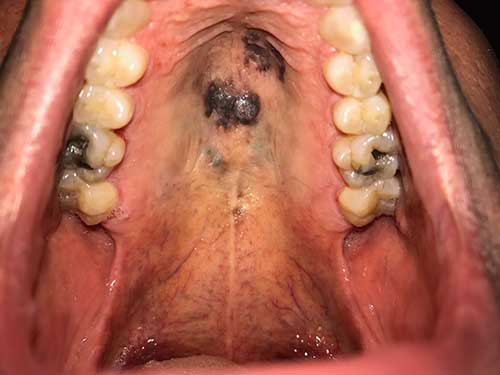
This is a 63-year-old male who presented to his dentist with the chief complaint of a palatal lesion that was of about one month’s duration.
Differential Diagnosis
1. Drug-associated hyperpigmentation
2. Systemic disease-associated hyperpigmentation (Addison’s disease)
3. Amalgam tattoo
4. Kaposi’s sarcoma
5. Mucosal malignant melanoma
Sorry, you are incorrect!
A differential diagnosis for a black palate includes two major categories: drug-associated hyperpigmentation and Mucormycosis-associated black palate.
Mucormycosis (Zygomycosis) is a deep fungal infection mainly affecting patients with uncontrolled diabetes and, to some extent, those who are immune compromised. Mucormycosis will not be considered on the differential diagnosis because the palatal mucosa is mostly intact but black in color. The black palate associated with Mucormycosis is usually perforated and ulcerated, a feature not present in this case. The histology is also not consistent with Mucormycosis.
With regard to drug-associated/induced hyperpigmentation, there are a number of drugs known to cause skin and mucosal hyperpigmentation as a secondary effect. The mechanism by which pigmentation is achieved varies from direct stimulation of the melanocyte activity to deposition of pigmented drug metabolites. Drugs that are known to cause mucosal and skin pigmentation include antimalarial agents (particularly chloroquine and hydroxychloroquine), antipsychotic medications (chlorpromazine), antibiotics (minocycline, tetracycline), cytotoxic drugs, birth control pills, AZT, ketoconazole, Gleevec and others. The pigmented lesions that are secondary to drug use can be localized or generalized. They can be small or large in size. The lesions are flat and range in color from tan to dark brown to blue, grey and greenish. This patient was not taking any of the medications that are known to cause hyperpigmentation. The histology was not consistent with drug-associated mucosal hyperpigmentation.
Sorry, you are incorrect!
Addison’s disease is an endocrine disorder affecting all ages and both sexes equally. It is characterized by muscle weakness, fatigability, nausea, vomiting, anorexia, weight loss, hypotension, and sometimes a craving for salt. It can also present with skin and mucosal darkening referred to as “bronzing,” which may affect the mouth at an early stage of the disease in the form of multiple flat and pigmented lesions. This patient only had these pigmented lesions on the hard palate; they were not present anywhere else. The patient was otherwise healthy, with no signs or symptoms of Addison’s disease. Neither the clinical presentation nor the histology of this case are consistent with Addison’s disease.
Sorry, you are incorrect!
Amalgam tattoo usually presents as a flat, irregular, uniformly dark-blue or black lesion localized to an area where an amalgam filling was/is present. It can also be present overlying an area where an amalgam retro fill is present at the apex of an endodontically treated tooth. Amalgam tattoos are more commonly present in tooth bearing areas of the alveolar bone. Less commonly, amalgam tattoos can also be raised and present in unusual sites such as the hard palate and for that reason they should be considered on the differential diagnosis. However, the palatal lesions in this patient are diffuse and lack uniformity in color, which makes a diagnosis of amalgam tattoo unlikely. The histology is also not consistent with amalgam tattoo.
Sorry, you are incorrect!
Kaposi’s sarcoma (KS) is more common on the palate and presents as isolated or coalescing purplish red spots. KS can be flat or nodular. All of these characteristics indicate that it is reasonable to include KS on the differential diagnosis since the palate in this patient has combined flat and slightly raised lesions. The color black however is unusual for the purplish-red KS. Additionally, KS in the United States is predominantly associated with AIDS. This patient has no such history. The histology in this case is not supportive of KS.
Congratulations, you are correct!
The patient has a history of malignant melanoma (MM) of the skin diagnosed in 2004; therefore, metastatic malignant melanoma should be on the differential diagnosis. A second primary malignant melanoma, although a rare occurrence, should also be placed on the differential diagnosis. The patient’s age, the size of the lesion, the site, and the clinical presentation of diffuse, irregular flat and raised lesions lacking uniformity in color are all consistent with the presentation of primary mucosal malignant melanoma. The etiology of oral MM is still unclear. Race appears to play a role; for example, 7.5% of all reported oral MMs affect those of Japanese descent and 10% affect those of Ugandan descent, compared with less than 1% of cases occurring in Caucasians. Cutaneous MM, however, occurs at a much higher rate in Caucasians than in those with brown or black skin, with a rate of 1:83 in whites compared to 1:1176 in dark-skinned individuals. Mucosal MMs are extremely rare compared to cutaneous melanomas; they constitute less than 2% of all MMs. For that reason, it is always prudent to first rule out metastatic MM to the oral cavity from the skin when a biopsy of an oral pigmented lesion confirms the diagnosis of MM. 80% of oral MMs affect the palate and the maxillary gingiva. The mandibular gingiva can be affected, but rarely. The site, age, clinical presentation is all consistent with that of malignant melanoma. There were no other lesions in or outside the oral cavity as confirmed by clinical examination including PET scan. The histology confirmed a diagnosis of malignant melanoma while the clinical findings confirmed it to be a second primary rather than metastatic disease.