Return to Case of the Month Archives
Left Facial Swelling with Mild Fever
Dolphine Oda, BDS, MSc
doda@u.washington.edu
Contributed by
Dr. James Reed, Edmonds, Washington
Case Summary and Diagnostic Information
A 47-year-old Hispanic male presented with left facial swelling, some episodic oral bleeding and a mild fever.
Diagnostic Information Available
This is a 47-year-old Hispanic male who was referred from the emergency department with left facial swelling. He complained of occasional oral bleeding in the past month, swelling for 5 days, bitter taste in his mouth and fevers of up to “99.0 F” (see case description for explanation) for which he took aspirin.
He has been human immunodeficiency virus (HIV) positive since 1993 and had been taking Videx, Epivir, Viread and occasionally Diflucan. He reported not seeing a dentist in 4 years because everyone was afraid of his HIV status. His past medical history is otherwise unremarkable.
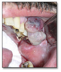
Clinical examination revealed mild trismus and a 2.0 x 1.5 x 1.5 cm purplish-blue exophytic, sessile mass involving the gingival margin of tooth #14 resembling a reactive gingival swelling such as peripheral giant cell granuloma or Kaposi sarcoma.
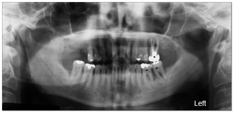
The panoramic radiograph demonstrated bone resorption near the right maxillary first molar and the area between the mandibular left first and second molars that resembled periodontitis. Teeth numbers 14, 18 and 19 had deep furcation probings and sulcular purulent exudate with 2+ mobility. A panoramic radiograph revealed significant bone resorption involving the furcation of teeth #s 14, 18 and some of 19.
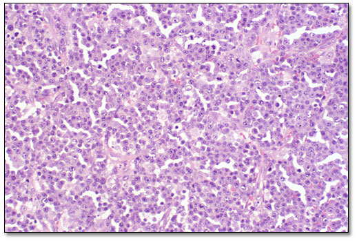
Histological examination revealed sheets of large cells with round and oval nuclei, some eccentrically arranged, some with central single prominent nucleoli. The histopathologic slide below is a low power (x100) view showing sheets of large cells, round to oval in shape with eosinophilic cytoplasm.
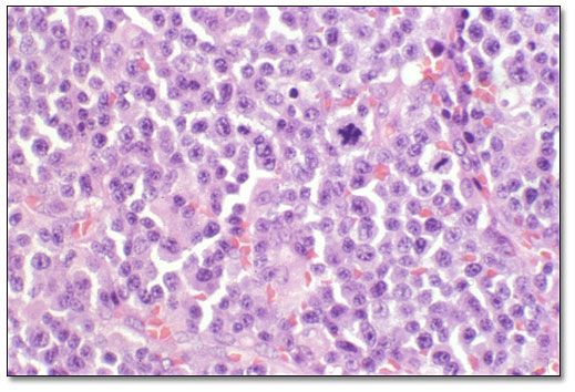
Mitotic activity and apoptosis were prominent as shown below using high power (x200) magnification. The slide shows abundant cytoplasm, vesicular nuclei, prominent nucleoli, some single and centrally located, macrophages with tingible bodies. Macrophages with tingible bodies were present, giving the specimen a “starry sky” appearance. Immunohistochemistry was positive for CD-138 and for Epstein Barr Virus.
After you have finished reviewing the available diagnostic information