Left Facial Swelling with Mild Fever
Can you make the correct diagnosis?
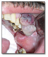
A 47-year-old Hispanic male presented with left facial swelling, some episodic oral bleeding and a mild fever.
Sorry! you are incorrect
Kaposi’s sarcoma (KS) is the most common AIDS-defining malignant neoplasm. It is of vascular origin and can be multifocal. KS in AIDS affects mostly young males and can be very aggressive in behavior. KS is described in 50% of AIDS patients. In the mouth, KS occurs most frequently in the palate and gingiva. It may present as flat and purplish red or as exophytic and nodular, making it very difficult to differentiate from malignant lymphoma in this case. It is usually asymptomatic, but may be painful. Human herpes virus #8 is often associated with KS. The only reason to rule out KS in this case is the histology (10).
Sorry! you are incorrect
Hemangioma is a benign hamartomatous proliferation of blood vessels. It occurs equally in males and females, principally in children, and is sometimes present at birth. The head and neck area is a common location; the tongue (macroglossia) is the most common location in the mouth, followed by the buccal mucosa and the lips. They present as flat or elevated, purplish-red lesions. They blanch on pressure. The location and age of the patient in this case are unusual for hemangioma. The histology is not supportive of a hemangioma.9
Congratulations! You are correct
Non-Hodgkin’s lymphoma, Kaposi sarcoma and cervical carcinoma are three AIDS-defining malignancies.1 Non-Hodgkin’s lymphoma is the second most common malignant neoplasm in AIDS patients, and up to 90% of certain subtypes of this disease are associated with Epstein Barr virus. Malignant lymphoma affects 5-10% of the HIV-positive population and consists predominantly of high grade B-cell type, Burkitt’s, immunoblastic and large cell lymphomas.2 Since the introduction of highly active antiretroviral therapy (HAART), the incidence of AIDS-related lymphoma has decreased from 6.2 to 3.6 per 1000 persons in the recent years6, 2 but it is clear that HAART alone can not prevent this aggressive disease from occurring1, 2 as it is still 338 times more common in AIDS patients than in the general population. 6
Plasmablastic lymphoma is an uncommon type of AIDS-related lymphoma and has recently emerged as a very aggressive subtype of large B-cell lymphoma. It can also occur in non-HIV patients, but rarely.4 It is of special interest to dental professionals, especially oral surgeons and periodontologists, because of its propensity for developing primarily in the oral cavity with subsequent expansion to other organs including the bone marrow.3 It was first described in 1997 by Delecluse et al. in 16 German patients, 15 of whom were HIV-positive.4 Since then, at least 14 more cases, all HIV-positive patients, have been reported: 12 from Italy7, and the other two from England and the United States.5,6 Most plasmablastic lymphomas of the oral cavity are Epstein Barr virus positive, as was this case.3,5 Histologically, this neoplasm has defined immunohistochemistry criteria that help separate it from other subtypes of the high grade lymphomas.4
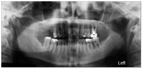
The most common locations for plasmablastic lymphoma are the gingiva, the palate, and the floor of the mouth. 4,7 It occurs most frequently in males between the ages of 25 and 60, the majority of whom are HIV positive. Of the total of 30 documented cases of this type of lymphoma, only three were in females (4,7). It can present as purplish flat or exophytic, and often simulates Kaposi sarcoma6 or a gingival reactive lesion such as a peripheral giant cell granuloma. It can also infiltrate and resorb the alveolar bone, as shown below
This is a very aggressive neoplasm with poor prognosis. In the study by Delecuse et al.4, nine out of sixteen died within 16 months, five were lost to follow-up and two were still alive at 8 and 18 months. The study by Gianluca et al.7, seven died within 1-16 months and three were alive after 26-54 months. The three patients who survived in this study were treated with combined HAART and CHOP (cyclophosphamide, adriamycin, vincristine and prednisone) or HAART and ACVBP (adriamycin, cyclophosphamide, vindesine, bleomycin and prednisone). Of the seven that died (7), all received CHOP or ACVBP alone. Radiation alone or in combination with multidrug chemotherapy was also used4,7, the latter for disseminated forms of this disease and the former for the more localized oral lesions. It is evident from the Gianluca’ s study7 that chemotherapy combined with HAART yields better results.
“Of clinical note the low grade temperature elevation may be overlooked as insignificant in immunocompetent patients. However, as in patients treated with chemotherapy, organ transplantation and high dose steroid administration the degranulation of mast cells, which initiates the elevation of temperature to produce fever, is suppressed. In these patients a small elevation in temperature may represent a significant systemic infection and should alert the vigilant clinician.” (From Dr. Reed)
In conclusion, we present a case in a 47-year old HIV-positive male that presented clinically as a gingival swelling. Histologically, this case was plasmablastic lymphoma, an AIDS-defining malignancy.
Treatment
The dental aspect of treatment included the removal of teeth numbers 14, 18 and 19 with an excisional biopsy of the gingival mass and the administration of Ampicillin 2.0gm intravenously. His postoperative regimen included Amoxicillin 500mg qid for 10 days. After one week, the surgical sites healed and the facial swelling subsided. He was referred to his hematologist for further medical workup to include CD4
counts and further medical management.
References
- Chetty R, Hlatswayo N, et al. Plasmablastic lymphoma in HIV+ patients: an expanding spectrum. Histopathology. 2003 Jun; 42(6): 605-9.
- Carbone A. Emerging pathways in the development of AIDS-related lymphomas. Lancet Oncol. 2003 Jan; 4(1): 22-9.
- Borrero JJ, Pujol E, et al. Plasmablastic lymphoma of the oral cavity and jaws. AIDS. 2002 Sep 27; 16(14): 1979-80. Delecuse HJ, Anagnastopoulos I, et al. Plasmablastic lymphomas of the oral cavity: a new entity associated with the human immunodeficiency virus infection. Blood. 1997 Feb 15; 89(4): 1413-20.
- Porter SR, Diz Dios P, et al. Oral plasmablastic lymphoma in previously undiagnosed HIV disease. Oral Surg Oral Med Oral Pathol Oral Radiol Endod. 1999 Jun; 87(6): 730-4.
- Flaitz CM, Nichols CM, et al. Plasmablastic lymphoma: an HIV-associated entity with primary oral manifestations. Oral Oncol. 2002 Jan; 38(1): 96-102.
- Gianluca G, Michaela C et al. Molecular histogenesis of plasmablastic lymphoma of the oral cavity. Br J Hematology. 2002 Dec; 119: 622-628
- Sapp JP, Eversole LR, et al. Contemporary Oral and Maxillofacial Pathology. 1997: 111-112.
- Sapp JP, Eversole LR, et al. 307-9.
- Leao JC et al. Human herpes virus 8 (HHV-8) and the etiopathogenesis of Kaposi’s sarcoma. Rev Hosp Clin Fac Med Sao Paulo. 2002 July-Aug; 57(4): 175-?
Sorry! you are incorrect
Peripheral giant cell granuloma (PGCG) is a reactive gingival lesion of periodontal ligament or periosteal origin. It occurs in young patients, with an average age of 30, and is more common in females (2:1). It occurs exclusively on the gingiva, especially anterior to the first molars. It presents as a sessile, red or red and blue lesion involving the buccal or lingual gingiva, sometimes surrounding teeth. It resorbs the underlying bone in a superficial and concave manner, creating a saucer-like depression. Clinically, this case can be mistaken for PGCG, but histologically the presence of sheets of round cells is not supportive of PGCG.8