All cases are discussed by: Dr. Dolphine Oda, UW-Oral Pathology Biopsy Service
Large multilocular expansile radiolucency: left posterior mandible
Contributed by: Dr. Eric Nordstrom
Oral & Maxillofacial Surgery, Anchorage, Alaska
Case Summary and Diagnostic Information
This is a 15-year-old male was referred to an oral for an incidental finding of a well-demarcated, expansile and multilocular radiolucency, left posterior mandible in area of teeth #s 17 and 18 extending partly into the ramus (Figure 1).
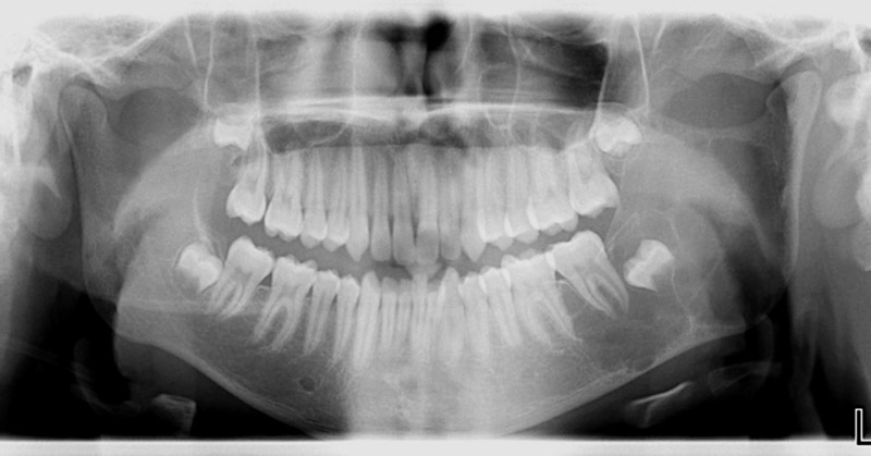
This is a 15-year-old male was referred to an oral for an incidental finding of a well-demarcated, expansile and multilocular radiolucency, left posterior mandible in area of teeth #s 17 and 18 extending partly into the ramus (Figure 1). The inferior border of the mandible is slightly eroded. There are no occlusal changes or nerve dysfunction.
Figure 1 This is a panoramic view taken at first clinical presentation demonstrating large, multilocular and expansile radiolucency of the left posterior mandible starting in area of tooth #18 extending posteriorly into the lower ramus and involving the not fully-developed tooth #17. Notice that both teeth #s 17 and 18 show mild, but not significant displacement.
The patient’s past medical history is unremarkable.
Patient was referred to an oral surgeon to evaluate an incidental clinical finding of a multilocular radiolucency in the posterior mandible of unknown duration. Teeth were not mobile and the inferior border of the mandible was slightly eroded (Figure 1).
Under local anesthesia, a lateral window aspiration was performed which yielded small amount of straw colored fluid, but otherwise almost totally empty. Small fragments of soft were curetted from the area for histologic evaluation. The cavity was described to be 4-5cm in diameter with few sinusoidal looking spaces that were purplish in color. The surgeon did not biopsy the purplish areas.
Histologic examination reveals multiple pieces of hard and soft tissue composed of lose and highly vascular granulation tissue containing giant cells and bone (Figures 2-4). Bone contains viable osteocytes and is at different stages of development. The granulation tissue is cellular and vascular with highly dilated and congested blood vessels. Multinucleated giant cells are present within the granulation tissue and few lining what appear as vascular spaces (Figures 2-3). This tissue contained large aggregates of surgical blood.
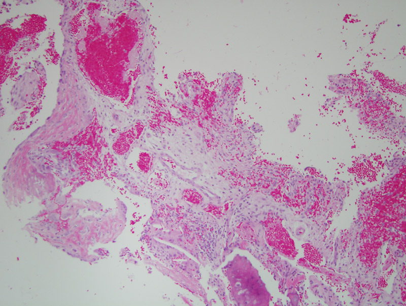
Figure 2 Low power (x40) H & E stained section. The histology shows small fragments of highly vascular and hemorrhagic granulation tissue and bone. The granulation tissue contains giant cells and dilated blood vessels and what appear as spaces with occasional giant cells at the rim.
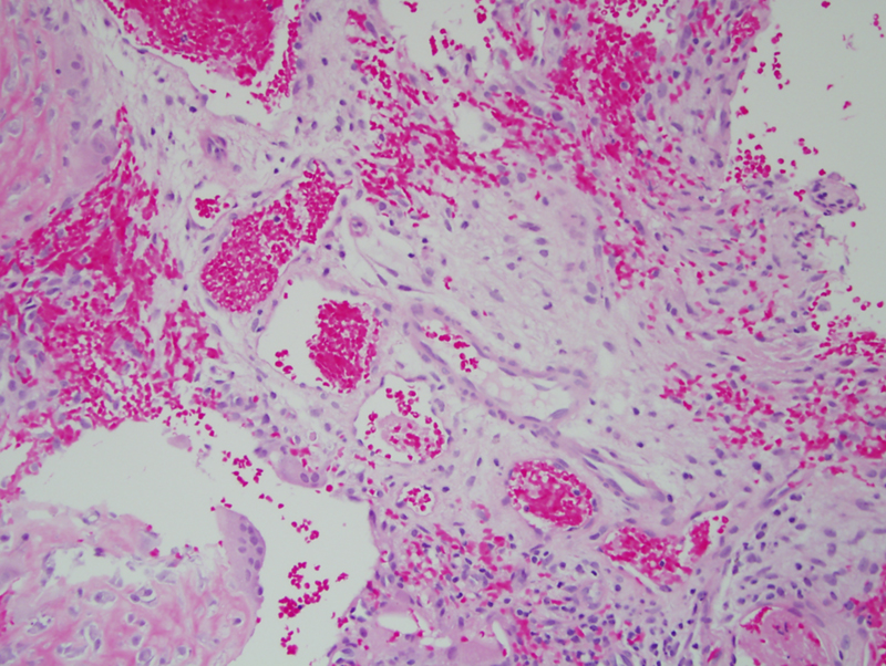
Figure 3 Higher power (x100) H & E stained section. This a higher power of the histology described in Figure 2.
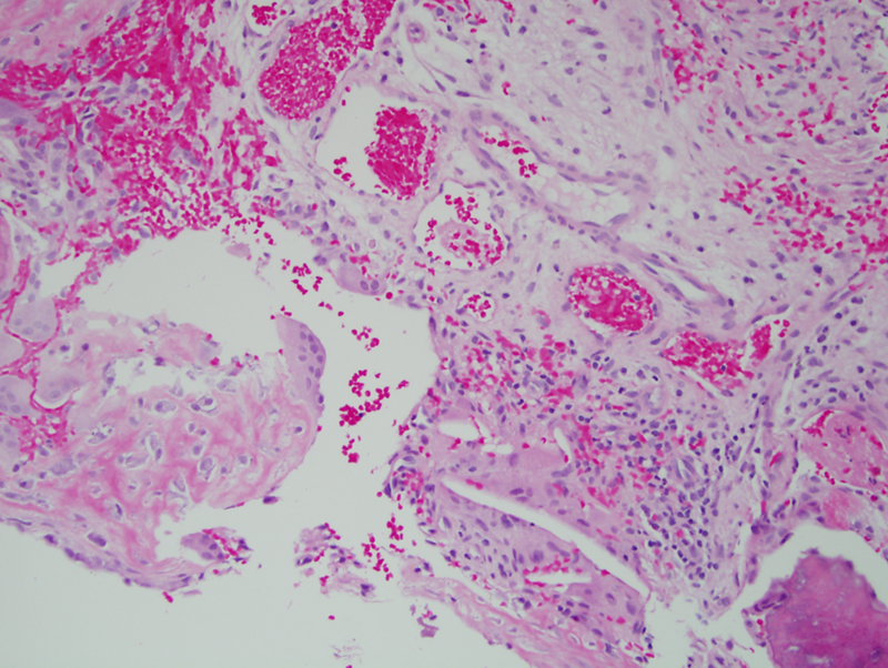
Figure 4 High power (x200) H & E stained section. This is a close look at the vascular granulation tissue with dilated and congested blood vessels. The granulation tissue contains giant cells.
After you have finished reviewing the available diagnostic information