Large radiolucency: Left posterior maxilla and maxillary sinus
Can you make the correct diagnosis?
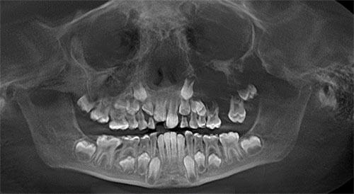
This is a 6-year-old female who was referred to a pediatric dentist for evaluation of a recently noticed left cheek swelling (Figure 1).
Sorry, you are incorrect!
The site, the lack of demarcation (diffuse pattern), large radiolucency, age and expansion into the maxillary sinus are all consistent with the clinical and radiographic findings of fibrous dysplasia (FD). Fibrous dysplasia occurs under 10 years of age especially in patients with Albright’s syndrome. Therefore the age and gender are appropriate for this condition especially in syndromic patients. The completely radiolucent lesion is unusual for FD since it tends to be mixed RL/RO with ground glass appearance. Early stages of FD can be completely radiolucent. The histology however is not consistent with FD.
The monostotic (one bone or bone complex area) type of FD constitutes approximately 80% of all fibrous dysplasia cases. FDs are expansile and disfiguring lesions, whether single or multiple. Monostotic FD, which involves the jaws, affects males and females equally. It occurs in childhood and at puberty and usually stops growing at age 30. It appears as an asymptomatic swelling of the maxilla or mandible; the maxillary lesion is the most common. It may involve bones other than the maxilla, including the zygoma, sphenoid and others. It is usually unilateral and is known to displace the teeth. The growth is usually slow, but rapid growth has been described, especially during puberty. The radiographic appearance, especially of the maxilla, is classically described as a ground glass appearance where fine radiopacity is noted. The mandibular lesions are much more deceptive because they tend to vary more ranging from cystic unilocular radiolucency to multilocular radiolucency to the classical ground glass radio-opacity.
Sorry, you are incorrect!
The site (maxilla), the age and gender and the radiographic findings of radiolucency pushing teeth apart are all consistent with the clinical behavior of central odontogenic fibroma. However, the lack of demarcation radiographically is not consistent with this neoplasm since this neoplasm tends to be well demarcated. The histology is not consistent with central odontogenic fibroma.
Central odontogenic fibroma is a rare neoplasm of mesenchymal odontogenic origin. Two types are described: Simple and WHO types both show a combination of connective tissue stroma with epithelial nests and calcified material simulating cementum globules. Central odontogenic fibromas occur most frequently in children and young females. The female-to-male ratio is 7:1 in some reports. It occurs most often in the maxilla anterior to first molar tooth. There are other reports that describe central odontogenic fibromas to be more common in individuals with average age of 40 and more common in the posterior mandible. These neoplasms are slow growing but can reach large sizes and swellings and can resorb and displace teeth. Radiographically, they can be unilocular or multilocular; expansile; and completely radiolucent or radiolucent/radiopaque (RL/RO) lesions. The histology in this specimen was only made up of lose connective tissue but no evidence of epithelial nests which was confirmed with immunohistochemistry to pancytokeratin. There was no evidence of tumor-formed calcified material such as cementum like globules.
Sorry, you are incorrect!
The large radiolucency with lack of demarcation, the displacement of teeth, the age and gender of this patient are all consistent with CGCG of the maxilla. The site however is not since CGCG is more common in the mandible, anterior to the first molar tooth. The histology is not supportive of CGCG.
Central giant cell granuloma is a non-neoplastic process that can occasionally behave in a very aggressive and expansile manner especially in children under ten years of age which is classified as the aggressive subtype of CGCG. Over 60% of CGCG cases occur in patients younger than 30 years of age, with twice as many occurrences in females as in males. Over 70% of cases occur in the mandible anterior to the first molar tooth.
Congratulations, you are correct!
The large swelling of the maxilla filling the maxillary sinus and displacing teeth is consistent with the behavior of odontogenic neoplasms that are benign but otherwise locally aggressive such as that of odontogenic myxoma. The site is not unusual for OM since it occurs almost equally in the maxilla and mandible with a small predilection for the posterior mandible. The age however is highly unusual since OM which rarely occurs under 10 years of age. The histology is that of odontogenic myxoma.
Odontogenic myxoma is not common; it usually occurs in jaw bones; in the tooth-bearing areas of the jaw. It is benign, but locally aggressive neoplasm. It has the potential for extensive bony destruction and extension into the surrounding structures.. Almost 75% of odontogenic myxomas occur in patients around 23-30 years of age with a slight female predilection (1:1.5 male-to-female ratio). It rarely occurs in patients over 50 or under 10 years of age. It occurs almost equally in the maxilla and mandible with a slight predilection for the posterior mandible. A few cases are described in the ramus and condyle, non-tooth bearing areas. Odontogenic myxoma is slow-growing, persistent and destructive. Most cases are expansile and can displace and resorb teeth. In the maxilla, they usually invade the maxillary sinuses and, in rare cases, cross the midline to the opposing sinus. Radiographically, the majority present as expansile and multilocular, though some are unilocular with or without scalloped borders, and rare cases present with a diffuse and mottled appearance which can be mistaken for a malignant neoplasm.