Single Purple-Red Swelling, Right Anterior Buccal Maxillary Gingiva
Can you make the correct diagnosis?
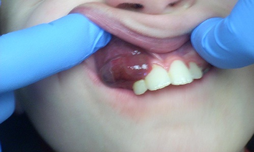
This 10-year-old white male was referred to the oral surgeon for the evaluation of a slowly growing, purple-red, smooth-surfaced and sessile gingival swelling in the anterior maxilla covering teeth #s 5-7.
Sorry! you are incorrect
The exophytic, sessile, purplish-red gingival swelling is consistent with the clinical presentation of a pyogenic granuloma. The color is more intense than is typical of PG; nonetheless, PG should stay on the differential diagnosis. The patient’s age and the location of occurrence are also supportive of a diagnosis of pyogenic granuloma, but not the patient’s gender. The histology, however, is not supportive of PG.
Pyogenic granuloma constitutes 85% of all reactive gingival swellings but can occur in areas other than the gingiva. It represents a profuse mass of vascular granulation tissue. It can be induced by trauma and local irritants such as excessive plaque, sharp fillings and dental calculus; it sometimes forms in an extraction socket in response to an irritant left in the socket. It can occur anywhere in the oral cavity and skin, especially the tongue, lips, fingers and nail beds. In the mouth, it occurs most commonly in the gingiva, especially the maxillary buccal and interproximal gingiva. Occasionally, it may surround the tooth. It is usually highly vascular, fast-growing, exophytic, lobular, sessile, and ulcerated or covered by pseudomembrane. The color changes from red to pink when it starts to heal. It occurs at any age and sex with a slight predilection for young females; it affects 1% of pregnant females. Pyogenic granuloma is usually painless except during eating, when bleeding and pain is described. Histologically, it presents as a mass of loose and vascular granulation tissue, usually with ulcerated or eroded surface epithelium and many inflammatory cells. A range of treatment modalities are available, including excision with removal of the local irritant, laser surgery, or intralesional injection with absolute alcohol, steroids or botulinum toxin. Scaling and polishing prior to surgical removal helps shrink the lesion. The prognosis is good, although recurrence is possible, especially during pregnancy.
Congratulations! You are correct
The intense purple-red color is supportive of a diagnosis of peripheral giant cell granuloma. PGCG constitutes the third most common gingival swelling, especially in this age group. However, it is more common in females. The histology is supportive of PGCG.
Peripheral giant cell granuloma constitutes fewer than 5% of all reactive gingival swellings. It consists of a hyperplastic mass of vascular granulation tissue with many osteoclast-like multinucleated giant cells. It presents as a lobular, purplish-blue exophytic nodule exclusively on the gingiva, both edentulous and dentate, and usually anterior to the molars. It originates from either the periodontal ligament or the periosteum. It occurs across a wide age range, especially in children, young adults, and females (2:1 female-to-male ratio). It presents as either sessile or pedunculated and smooth-surfaced or lobular. Though usually painless, it can occasionally be ulcerated, painful and accompanied by bleeding. Like pyogenic granuloma, it is usually present either on the buccal or lingual gingiva or between teeth, but it can occasionally surround the teeth and act aggressively by displacing teeth much like a sarcoma. It can also resorb the underlying bone in a smooth and concave “saucer-like” manner. Complete excision including curettage of underlying bone is the preferred treatment. It has a good prognosis with a recurrence rate of approximately 10%.
References
- Fantasia JE, Damm DD. Red nodular lesion of tongue. Pyogenic granuloma. Gen Dent. 2003 Mar-Apr;51(2):190, 194.
- Ichimiya M, Yoshikawa Y, Hamamoto Y, Muto M. Successful treatment of pyogenic granuloma with injection of absolute ethanol. J Dermatol. 2004 Apr;31(4):342-4.
- Pham J, Yin S, Morgan M, Stucker F, Nathan CA. Botulinum toxin: helpful adjunct to early resolution of laryngeal granulomas. J Laryngol Otol. 2004 Oct;118(10):781-5.
- Flaitz CM, Peripheral giant cell granuloma: a potentially aggressive lesion in children. Pediatr Dent. 2000 May-Jun;22(3):232-3.
- Chaparro-Avendano AV, Berini-Aytes L, Gay-Escoda C. Peripheral giant cell granuloma. A report of five cases and review of the literature. Med Oral Patol Oral Cir Bucal. 2005 Jan-Feb;10(1):53-7; 48-52.
- Neville BW, Damm DD, Allen CM, Bouquot JE. Peripheral giant cell granuloma. In: Oral and Maxillofacial Pathology, 2nd edition. Philadelphia: W.B. Saunders, 2002. p. 449-451.
- Chan YC, Giam YC. Guidelines of care for cutaneous haemangiomas. Ann Acad Med Singapore. 2005 Jan;34(1):117-23.
- Whang KK, Cho S, Seo SL. Excision of hemangioma and sculpting of the lip using carbon dioxide laser. Dermatol Surg. 2004 Dec;30(12 Pt 2):1601-2; author reply 1602.
- Benedetto AV. News in treatment of angiomas. J Eur Acad Dermatol Venereol. 2004 Mar;18(2):122-3.
- Lambrecht JT, Stubinger S, Hodel Y. Treatment of intraoral hemangiomas with the CO2 laser. Schweiz Monatsschr Zahnmed.
2004;114(4):348-59.
Sorry! you are incorrect
The purple-red color of the lesion and the age of the patient certainly suggest the possibility of a hemangioma. Therefore, hemangioma should be included on the differential diagnosis. However, hemangiomas rarely occur on the gingiva. The histology is not supportive of this condition.
Hemangioma is a family of benign developmental vascular anomalies occurring at infancy. They progress through two stages of growth: a rapid growth phase followed by an involution phase. Vascular lesions are most common in infants. Lymphangiomas occur at birth and progress with age, while hemangiomas typically occur a few weeks after birth and continue to grow rapidly for the first year; they stop growing and begin to involute within the subsequent few years. Hemangiomas are benign proliferations of blood vessels with many classifications; capillary and cavernous hemangiomas are the most common types. Capillary hemangiomas affect 1% of all newborns in the United States. Half of all hemangiomas occur in the head and neck area, especially the tongue. They are the most common cause of macroglossia. They can also occur on the buccal mucosa and lips. In addition, hemangioma has a slight predilection for occurrence in females. The lesion can present as flat or exophytic, smooth-surfaced or lobular, and localized or diffuse; though usually single and localized, it can also present in multiples. Superficial hemangiomas are bright red, while the deep lesions are purplish-red in color; they blanch on pressure unless thrombosed. The vast majority will regress and resolve within the first ten years of age. Treatment of hemangioma depends on its size, its relationship to other anatomical structures and the rate of blood flow. Observation is important since many spontaneously involute, especially capillary hemangiomas. If a hemangioma persists, local topical application of injections with corticosteroids has a 75% success rate of involution within two weeks to two months post-injection. Several complications are described with local and systemic use of steroids; interferon alfa-2a has also been successfully used. Surgical procedures include excisional scalpel surgery for smaller lesions and laser removal for larger lesions. Laser use includes CO2. Argon and other types of laser have also been used with variable rates of success. The prognosis depends on the size and whether it is a soft tissue or bony lesion; it can range from good to extremely poor with gross facial deformity and compromised function.