Single Purple-Red Swelling, Right Anterior Buccal Maxillary Gingiva
Dolphine Oda, BDS, MSc
doda@u.washington.edu
Dr. Alan Sato
Oral & Maxillofacial Surgery, Lynnwood, WA
Case Summary and Diagnostic Information
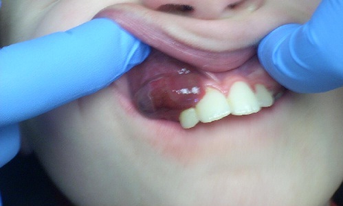
This 10-year-old white male was referred to the oral surgeon for the evaluation of a slowly growing, purple-red, smooth-surfaced and sessile gingival swelling in the anterior maxilla covering teeth #s 5-7.
Diagnostic Information Available
This 10-year-old white male was referred to the oral surgeon for the evaluation of a slowly growing, purple-red, smooth-surfaced and sessile gingival swelling in the anterior maxilla covering teeth #s 5-7 (Figure 1). This growth was of six months’ duration but was reported to have suddenly increased in size over the last few weeks. It was isolated and otherwise asymptomatic. The associated teeth were vital and there was no evidence of bone resorption.
Figure 1. This photograph was taken at first presentation where a large, dome-shaped, smooth-surfaced and ulcerated purplish-red swelling is identified buccal to teeth #s 5-7.
The patient’s past medical history is unremarkable.
This was an isolated lesion slowly growing over a six-month period but it had suddenly increased in size over the last few weeks. The associated teeth were vital and showed no evidence of displacement.
The lesion was completely excised under local anesthesia. The specimen was removed in one piece (Figures 2 & 3).
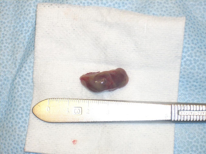
Figure 2. This photograph is of the gross specimen removed in one piece demonstrates a dark-brown, smooth soft tissue lesion.
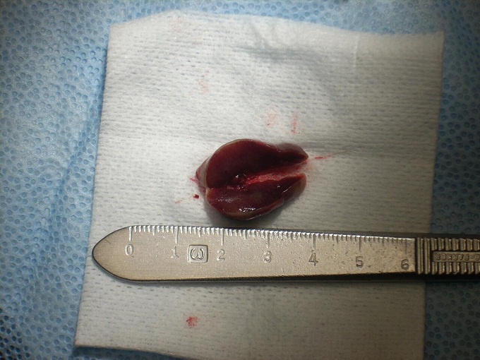
Figure 3. This photograph is of the gross specimen removed in one piece and bisected to demonstrate a dark-brown, smooth soft tissue lesion simulating a section of a liver.
Excisional Biopsy
Histologic examination reveals a multisected piece of soft tissue embedded in four separate blocks. All specimens are made up of surface epithelium with an underlying mass of granulation tissue containing numerous giant cells (Figures 4-6). The granulation tissue mass occupies the bulk of the specimen and is loose and vascular with extravasated erythrocytes and clusters of hemosiderin pigment. The giant cells are of variable shapes and sizes and are haphazardly arranged (Figures 5 & 6).
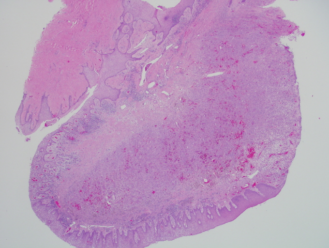
Figure 4. Low power (x40) H & E stained histology shows an ulcerated mass of vascular granulation tissue containing numerous multinucleated giant cells.
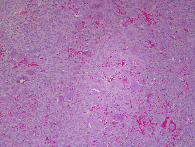
Figure 5. Higher power (x200) H & E stained histology shows a mass of vascular granulation tissue with numerous multinucleated giant cells and extravasated erythrocytes.
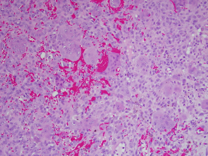
Figure 6. High power (x200) H & E stained histology shows a closer look at the vascular granulation tissue with numerous multinucleated giant cells and extravasated erythrocytes.
After you have finished reviewing the available diagnostic information