Large, rough red & white lesion left posterior buccal mucosa/vestibule
Can you make the correct diagnosis?
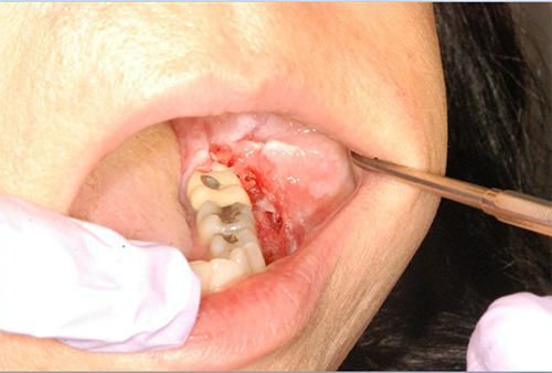
This is a 51-year old white female who described her condition as follows: “There is a painful cyst on my gums on the lower left.”
Sorry, you are incorrect!
The only reason why lichen planus (LP) is considered on the differential diagnosis of this case is that the patient has a past medical history of having had LP for fifteen years. The clinical presentation of an isolated papillary and ulcerated lesion on the buccal mucosa and vestibule is not consistent with any type of lichen planus, including the hyperplastic subtype. Hyperplastic or plaque LP is uncommon and presents as a confluent white plaque that is often mistaken for leukoplakia. The hyperplastic type is more common on the dorsal surface of the tongue, gingiva, and palate, making a clinical diagnosis of LP difficult at times, especially if the patient is a smoker. It does not present as a papillary red and white and ulcerated lesion. The histology is also not consistent with lichen planus of any type.
Sorry, you are incorrect!
The localized papillary clinical presentation raises the possibility of a reactive hyperplastic epithelial or connective tissue process with papillary/verrucoid configuration. However, the buccal mucosa-vestibule site is unusual since IPH most often presents in the hard palate under dentures worn 24 hours per day without proper cleaning. Inflammatory papillary hyperplasia is a reactive response to a local irritant. It is typically reported under dentures but can develop in response to other types of irritants. Since it occurs at any age and with equal gender distribution, this condition cannot be excluded based on age and gender or on the clinical presentation of a white to red, pebbly lesion. Candidal infection has been implicated in IPH. The hard palate is the most common location. It is also seen in dentate patients who habitually lick their palate, breathe through their mouth or have a high vaulted palate.
Sorry, you are incorrect!
The age, site and the clinical presentation of verrucoid/papillary surface is consistent with the clinical presentation of verrucous carcinoma, as is the linear spread of the lesion. However, this condition is more common in males with a history of chronic tobacco use which this patient does not report. The histology is not supportive of verrucous carcinoma.
Sorry, you are incorrect!
The clinical presentation of a rough and papillary surface raises the possibility of some type of granulomatous condition. A number of etiologies can lead to granulomatous diseases manifesting in the oral cavity, most of which are rare occurrences. These include infectious diseases such as deep fungal and bacterial infection, immune-mediated conditions such as Crohn’s disease, foreign body-induced granulomas, and rare conditions of unknown etiology such as Wegener’s granulomatosis.
The patient’s past medical history rules out Crohn’s and Wegner’s. The clinical presentation also rules out Wegner’s since the latter occurs more often on the gingiva with the appearance of “strawberry” gingivitis. Geographically, histoplasmosis can be ruled out since the patient is from the Pacific Northwest and has not travelled to the Ohio-Mississippi Valley area, and therefore has not been exposed to the fungal organism causing this disease. The patient is not immune compromised and there is no history of persistent coughing and weight loss. Foreign body granuloma cannot be ruled out based on clinical findings and medical history, so it should remain on the differential diagnosis. The histology, however, is negative for any type of granulomatous condition afflicting the mouth.
Congratulations, you are correct!
Today, it is well known that oral squamous cell carcinoma is a disease with many etiologies. Most are associated with chronic tobacco use, especially cigarette smoking; the next most common causes are HPV, lichen planus, and proliferative verrucous leukoplakia (PVL). Neither the site of occurrence nor the patient’s medical history is consistent with SCC caused by chronic tobacco use. The site, the verrucoid red and white clinical presentation, and the patient’s age and gender are all consistent with papillary SCC arising in PVL, but the patient lacks a clinical history of recurring, persistent and progressively evolving oral white patches. This is an unlikely presentation for HPV-associated oral SCC since the latter occurs more often in the tonsillar area and posteriorly, and is typically seen in white males under the age of 50. As for oral SCC arising in lichen planus, the patient’s history of having had LP for 15 years lends itself to that diagnosis. Therefore, the site, the age, and the gender are all consistent with SCC arising in LP. The histology confirmed that this is squamous cell carcinoma. The histology, however, was very small; therefore, we were unable to support the diagnosis of oral SCC arising in LP.