All cases are discussed by: Dr. Dolphine Oda, UW-Oral Pathology Biopsy Service
Radiopaque mass of the chin, right side
Contributed by:
Dr. Michael O’Neil
Edmonds Oral Surgery, Edmonds, WA
Case Summary and Diagnostic Information
This is a 60-year-old male who presented with a hard nodule in the right side of the chin area. The lesion had been present for many years and was slowly enlarging in size but otherwise was not symptomatic.
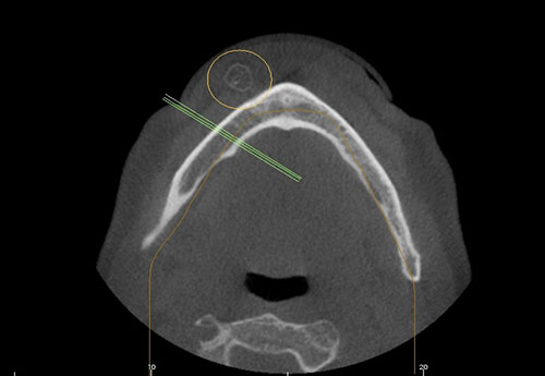
This is a 60-year-old male who presented with a hard nodule in the right side of the chin area. The lesion had been present for many years and was slowly enlarging in size but otherwise was not symptomatic. The patient reported trauma—specifically, he said that he fell on this area of the chin many years ago. Per CBCT, the lesion is 1.3 X 1.1 cm at its greatest dimensions. The CBCT findings show a circular rim of radiopacity with a translucent center (Figure 1). When surgically exposed, a smooth surfaced and well-circumscribed yellow nodule emerged (Figure 2).
Figure 1 This is an axial image of the area by CBCT taken at presentation. Note the radiopaque ring in the right side of the chin.
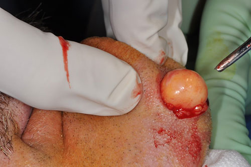
Figure 2 This photograph is taken during the early part of incision making in the area. Note the smooth-surfaced yellow nodule emerging as a whole from the surgical site.
The patient’s past medical history is significant for hypertension and for an allergic reaction to penicillin. He is otherwise healthy.
The patient reported trauma to this area many years ago. The swelling in this area was slowly growing, but was otherwise asymptomatic. The nodule was hard to palpation and well circumscribed (Figures 1 & 2).
Under IV sedation and local anesthesia, the lesion was excised via submental skin approach. Upon performing the skin incision, a smooth-surfaced yellow nodule emerged from the area (Figure 2). The specimen was submitted for histologic evaluation.
Histologic examination reveals a decalcified and multisected piece of hard and soft tissue composed of sheets of mature adipose tissue partially encapsulated and circumscribed by a ring of bone (Figures 3-5). The bone was viable and lamellar in type surrounded by periosteum (Figure 4), layers of connective tissue (Figure 5) and by sheets of mature adipose tissue (Figures 3-5). The adipose tissue is made-up of lobules of adipocytes suspended on delicate collagen fibers and small blood vessels (Figure 6).
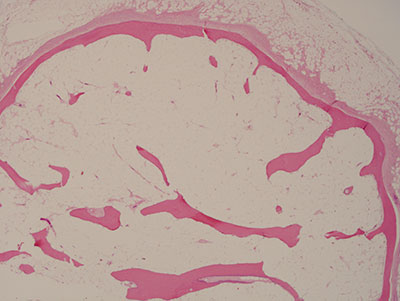
Figure 3 Low power (x40) H & E stained section which shows a ring of bone surrounded by and surrounding sheets of mature adipose tissue.
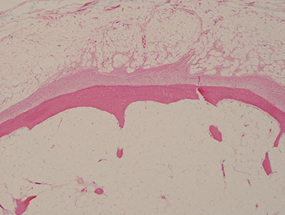
Figure 4 Low power (x100) H & E stained section with a closer look at the viable bone surrounded by layers of periosteum.

Figure 5 Higher power (x200) H & E stained section showing the rim of viable bone, layers of periosteum and mature adipose tissue at closer magnification.
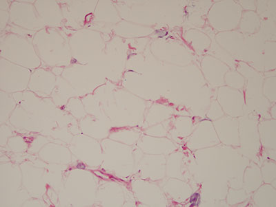
Figure 6 Higher power (x200) H & E stained section showing the mature adipocytes suspended on delicate collagen fibers and small blood vessels.
After you have finished reviewing the available diagnostic information