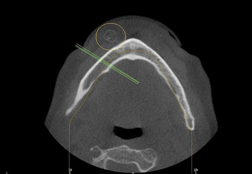
August 2019: Radiopaque mass of the chin, right side
Can you make the correct diagnosis?
This is a 60-year-old male who presented with a hard nodule in the right side of the chin area. The lesion had been present for many years and was slowly enlarging in size but otherwise was not symptomatic.
Sorry, you are incorrect!
The site being on the face (chin area); the smooth and dome-shaped nodule when combined with the clinical behavior of slow-growing and asymptomatic are all consistent with the clinical presentation of an epidermal cyst. The age however is on the older side of the spectrum since epidermal cysts tend to occur in young and middle aged individuals with no sex predilection. The radiopaque ring noted radiographically is not typical of epidermal cyst but these cysts can become calcified. The color yellow is not typical of this cyst but a sac full of keratin can reflect yellow. The one point that argues against epidermal cyst is the lack of punctum which is usually present on the surface of these cysts. The histology is not consistent with epidermal cyst.
Sorry, you are incorrect!
The single slow-growing firm to hard nodule on the face and the ossifications revealed by the radiographic imaging are features consistent with the clinical presentation of pilomatrixoma. The age and gender however are not. Neither is the color yellow. Pilomatrixoma tends to be more chalky white in color-not yellow. Pilomatrixomas are usually isolated lesions that tend to occur more on the face, especially the cheek but can occur on the head and neck area in general as well as the limbs and trunk. About 60% of the cases occur under 20 years of age, mostly under 10 years of age. Pilomatrixomas are more common in individuals around 50-60 years of age. The histology is not consistent with pilomatrixoma.
Sorry, you are incorrect!
The radiographic features of a ring of radiopaque lesion combined with the clinical history of trauma to the area raises the question of myositis ossificans (MO). About 50% of MO lesions are associated with trauma but is usually associated with repetitive trauma to the area. One time trauma to the area causing MO is reported. The site however argues against MO since these lesions occur in large muscle bundles such as the quadriceps. The age of the patient also argues against MO age range of 20-40. The gender is consistent with MO since it is more common in males (2:1 ratio). Myositis ossificans is a benign and reactive process where injury to muscle and surrounding soft tissue can lead to metaplastic bone formation. Large leg muscles are most commonly affected but MO is described in small muscles such as the masticatory muscles which can lead to trismus and pain. The chin area muscle is a very unusual site for MO. The smooth surfaced yellow nodule emerging from the incision is not consistent with MO. The histology is not consistent with MO.
Sorry, you are incorrect!
The age, gender and site are all consistent with Osteoma cutis since this lesion can occur at any age with no gender predilection. This lesion is exceedingly rare and is usually single but can be multiple. Histologically, Osteoma cutis is made-up of a nodule of lamellar viable bone with marrow spaces. The histology is not consistent with Osteoma cutis.
Congratulations, you are correct!
The age, the slow-growing & asymptomatic lesion, the age when combined with the surgical presentation of a yellow-smooth-surfaced nodule emerging from the surgical site are consistent with the clinical presentation of a lipoma. The gender and the site/chin however argue against lipoma of the skin. Lipomas are the most common connective tissue benign neoplasms that more commonly occur in females around middle age or older individuals. The most common sites for lipomas are the trunk, abdomen and neck. It is rare on the face. They are slow-growing and painless. Histologically, lipoma can assume a variety of morphologies including those associated with cartilage and bone formation.
In general, lipomas with ossification are rare, but ossification is described in patients that give history of trauma to the area which is the case in this patient. Although rare, lipomas with ossification are reported in the head and neck area in individuals around 50-years of age. They can be present for many years with a range of 1-20 years which is also true in this patient. The histology is of lipomas with ossification is typically composed of mature adipose tissue interspersed with spicules of mature and viable bone. The histology in this case is that of a lipoma with a ring of viable bone.