All cases are discussed by: Dr. Dolphine Oda, UW-Oral Pathology Biopsy Service
Gingival swelling: lingual gingiva between teeth #s 27 & 28
Contributed by: Dr. Daniel Brady
Oral & Maxillofacial Surgery, Everett, WA
Case Summary and Diagnostic Information
This is a 16-year-old male who presented with a large smooth-surfaced pink gingival swelling with focal ulceration interproximal and lingual to teeth #s 27 & 28 (Figure 1).
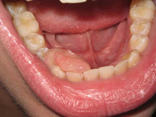
This is a 16-year-old male who presented with a large smooth-surfaced pink gingival swelling with focal ulceration interproximal and lingual to teeth #s 27 & 28 (Figure 1). It was asymptomatic but the large size pushed the crown of teeth apart causing a diastema (Figures 2A & 2B). The large size also interfered with the tongue movement which was “annoying” to the patient. This lesion is reported to have started four months ago and grew fast in the last month. It is grossly measured as 2.5 X 1.4 X 0.8 cm. The patient was referred to an oral surgeon for the removal of this lesion.
Figure 1 This photograph is taken at the first clinical presentation. The figure shows a large smooth surface, sessile, pink swelling measuring 2.5 X1.4 X0.8 cm in greatest dimensions. It is interproximal and lingual to teeth #s 27 & 28.
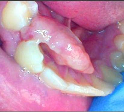
Figure 2A This photograph is taken at the first clinical presentation. The figure shows the swelling mildly pushing the crowns of teeth #s 27 & 28 apart creating a diastema.
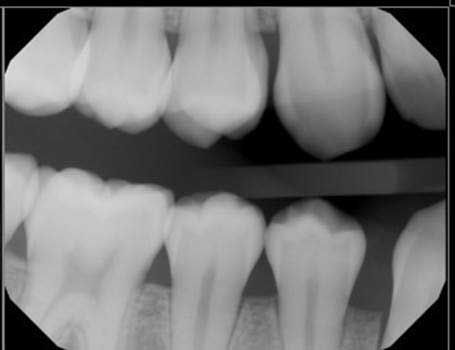
Figure 2B This bitewing radiograph was taken at the first clinical presentation. The radiograph shows teeth #s 27 & 28 mildly pushed apart.
The patient’s past medical history is significant for asthma controlled with albuterol.
As described earlier this lesion is of four month duration. It was slowly growing in the first three months but changed to fast growth in the last month. It was not painful but was mildly pushing the crowns of teeth #s 27 and 28 apart (Figures 2A & 2B) causing a diastema.
Under local anesthesia, the lesion was excised in total using blade # 15 down to bone.
Histologic examination a large and multisected piece of soft tissue composed of ulcerated surface epithelium with an underlying mass of moderately cellular fibrous connective tissue containing calcified bone at different stages of development (Figures 3-6). In some areas, bone is calcified (Figures 3-5) and in others areas, the bony trabeculae are at very early stage of development (Figure 6). All the bony trabeculae have viable osteocytes and osteoblastic rimming.
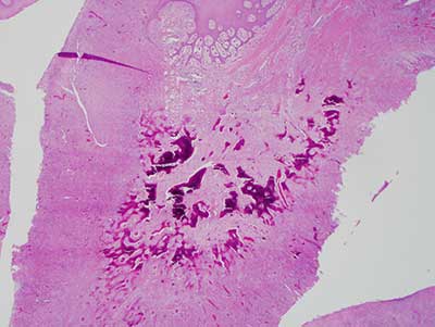
Figure 3 Low power (X40) H & E stained section revealing a low-power image of an ulcerated and moderately cellular connective tissue mass containing calcified material.
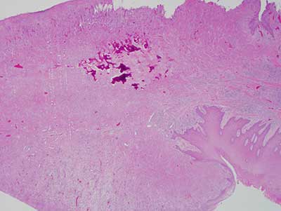
Figure 4 Higher power (X40) H & E stained section revealing another low-power image of the ulcerated moderately cellular connective tissue mass containing calcified material.
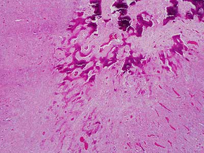
Figure 5 Higher power (X100) H & E stained section revealing a closer look at the ulcerated connective tissue mass demonstrating that the calcified material to be bone. It is in form of trabeculae with viable osteocytes and osteoblastic rimming.
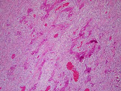
Figure 6 Higher power (X100) H & E stained section revealing a closer look at bony trabeculae that are at a very early stage of development.
After you have finished reviewing the available diagnostic information