Gingival swelling: lingual gingiva between teeth #s 27 & 28
Can you make the correct diagnosis?
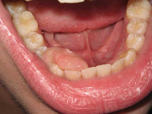
This is a 16-year-old male who presented with a large smooth-surfaced pink gingival swelling with focal ulceration interproximal and lingual to teeth #s 27 & 28 (Figure 1).
Congratulations, you are correct!
The exophytic, pink, sessile and firm nature of this lesion are all consistent with those of a benign and reactive gingival swelling responding to local stimuli such as dental calculus and plaque. The most common reactive gingival swellings include pyogenic granuloma (PG), inflammatory fibrous hyperplasia (IFH), peripheral ossifying fibroma (POF) and peripheral giant cell granuloma (PGCG). Pyogenic granuloma and peripheral giant cell granuloma tend to be clinically red (PG) and grey to purplish red (PGCG) and therefore will not be considered on the DDX list. IFH is considered mainly because it clinically presents at any age and gender and is usually exophytic, firm, sessile, smooth surfaced, and pink like the surrounding gingiva if it is not ulcerated. The histology however argues against inflammatory fibrous hyperplasia.
Peripheral ossifying fibroma (POF) is favored over all other reactive gingival swellings mainly because the color is pink like the color of the surrounding gingiva. POFs are frequently associated with surface ulceration. The age is consistent with this condition but not the gender. The site is not consistent with POF being between and lingual to the premolars while POFs tend to occur more commonly on the buccal gingiva in the anterior jaws and less commonly in the lingual or interproximal gingiva.
POFs tend to present on the anterior gingiva of both jaws in form of a sessile firm nodule, often ulcerated, but sometimes smooth surfaced and pink as is the case with this patient minus the small ulceration. POFs are more common on the buccal gingiva but can occur on the lingual (palatal) gingiva or between teeth. This case partially involves the interproximal gingiva but is mostly lingual. They are more common in young females, occurring in females at a ratio of 3:2 with a typical age range of 10-20 years. The clinical presentation and the histology are consistent with POF.
Sorry, you are incorrect!
Exophytic, sessile, pink and firm gingival swellings may also be benign neoplasms of tooth (odontogenic) origin that solely occur on the gingiva. It is however important to note that peripheral odontogenic neoplasms are rare. The clinical presentation of an exophytic, sessile, pink, and smooth-surfaced swelling on the posterior mandibular gingiva is consistent with the clinical presentation of peripheral ameloblastoma. The age is not consistent with peripheral ameloblastoma but the gender is.
Almost 99% of ameloblastomas are intra-osseous (central/within bone) and about 1% are present completely outside the bone, usually on the gingiva known as peripheral ameloblastomas. They are slow-growing and non-aggressive compared to the solid and desmoplastic intra-osseous ameloblastomas. They present as small, well-circumscribed, firm nodules on the gingiva, mainly of the posterior mandible in patients around 50 years of age. They are slightly more common in males. They are usually asymptomatic, pink like the surrounding mucosa, smooth surfaced, and sessile.
The histology in this case is not consistent with peripheral ameloblastoma or any other peripheral odontogenic neoplasms.
Sorry, you are incorrect!
In general, benign soft tissue neoplasms such as solitary neurofibromas and leiomyomas are rare in the mouth. If they do occur, the gingiva is not a common site. On the rare occasions that leiomyomas occur in the mouth, they tend to be of the vascular type (angiomyoma). Vascular leiomyomas are slightly reddish-gray in color and they, too, rarely occur on the gingiva.
Solitary neurofibromas (NF) of the oral cavity can occur between 20 and 30 years of age. Some studies suggest that NFs are slightly more common in females, while others report an equal gender predilection. They usually occur on the buccal mucosa, tongue, palate and, rarely, on the gingiva. They are non-tender, smooth surfaced, slow-growing swellings that may be soft or firm. The age, gender and site are not consistent with solitary NF and neither is the histology.
Leiomyomas of the oral cavity tend to originate from the smooth muscle cells around blood vessels. These are known as vascular leiomyomas (or angiomyomas) and account for 75% of oral leiomyomas. Patients tend to be over 30 years of age and are predominantly males. They more commonly occur on the posterior tongue, palate, cheeks and lips. They are slow growing, painless, sessile or pedunculated; smooth surfaced, and normal colored or slightly bluish nodules. The site, age and color are not consistent with leiomyoma and neither is the histology.
Sorry, you are incorrect!
The clinical presentation of an isolated, slow-growing gingival lesion in an otherwise healthy 16-year old male argues against a malignant neoplasm be it primary or metastatic. However, one cannot definitively know until the lesion is histologically evaluated. The histology is not consistent with a malignant neoplasm, primary or metastatic.