Left maxilla: mixed RL/RO swelling
Can you make the correct diagnosis?
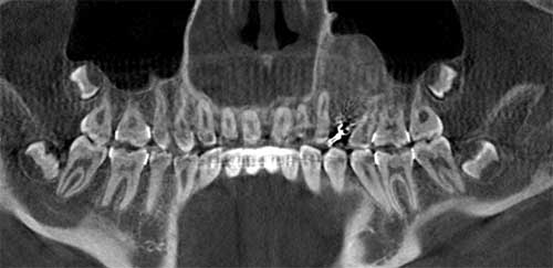
This is a 13-year-old female who was referred in 2015 for the management of a palatally impacted tooth #12.
Differential Diagnosis
1. Fibrous dysplasia
2. Chronic sclerosing osteomyelitis
3. Juvenile ossifying fibroma
4. Central odontogenic fibroma
5. Benign osteoblastoma
Sorry, you are incorrect!
The age, site, clinical presentation of expansion, and the mixed radiolucent (RL)- radiopaque (RO) radiographic appearance are all consistent with the clinical and radiographic presentation of fibrous dysplasia (FD). Therefore, FD should be considered highly on the differential diagnosis. However, the well-circumscribed radiographic presentation is not typical of FD since they tend to blend in with the surrounding bone. The histology is not consistent with FD.
Sorry, you are incorrect!
Given the two-year history of growth, the exposed alveolar bone, and the orthodontic manipulation of the tooth into the alveolar ridge, one must consider potential infection and osteomyelitis in the area. Therefore, chronic sclerosing or chronic osteomyelitis should be considered on the differential diagnosis. However, the clinical history is negative for pain or infection in the area while the tooth was being guided into alveolar ridge.
Chronic sclerosing osteomyelitis can occur at any age, gender, or site, but it tends to occur more often in adults and in the mandible. A rare variant that can occur in children, “juvenile mandibular chronic osteomyelitis,” usually affects females 6-12 years of age and so far has been reported mainly in the mandible. The well-circumscribed radiographic features of this lesion are not consistent with osteomyelitis, which tends to have more ill-defined margins. The histology is not consistent with osteomyelitis, whether chronic or sclerosing.
Congratulations, you are correct!
The age, site, clinical expansion, and radiographic findings are all consistent with the characteristics of juvenile ossifying fibroma. The histology is also consistent with that condition with predominant features favoring the psammamatoid histologic variant.
Sorry, you are incorrect!
The site, age, and gender should bring to mind central odontogenic fibroma. The radiographic findings, especially that of a well-circumscribed lesion, are also consistent with COF, but the level of radiopacity is not. Central odontogenic fibromas tend to be more radiolucent; if radiopacity is present, it tends to have a speckled rather than a homogeneous appearance. The histology in this case is not consistent with COF.
Sorry, you are incorrect!
The age, clinical expansion, and radiographic features are all consistent with osteoblastoma, but not the gender or the site. Osteoblastomas tend to be more common in males and in the posterior mandible.