All cases are discussed by: Dr. Dolphine Oda, UW-Oral Pathology Biopsy Service
Single deep ulcer, lateral tongue
Contributed by: Dr. John Malan
Oral & Maxillofacial Surgery-Montana
Case Summary and Diagnostic Information
This is a 65-year old white male who was referred by his dentist to an oral surgeon for the evaluation of a single deep ulcer on the right lateral/ventral border of tongue.
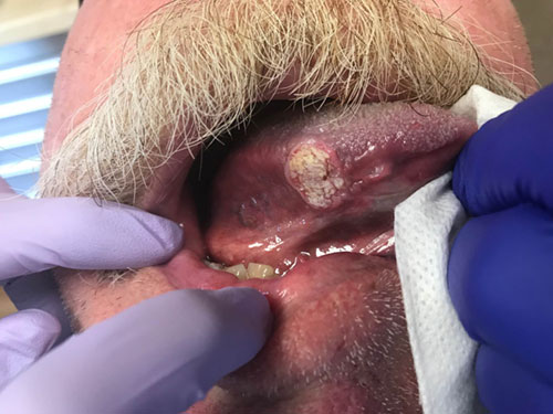
This is a 65-year old white male who was referred by his dentist to an oral surgeon for the evaluation of a single deep ulcer on the right lateral/ventral border of tongue (Figure 1). The duration is that of “several months.” The ulcer is described to be approximately 1.5 X 1.5 cm in greatest dimensions. This patient has history of chronic cigarette smoking one pack per day for approximately 50 years. The lesion is largely asymptomatic unless accidentally traumatized while chewing.
Figure 1 This photograph was taken at first clinical presentation. Note the large, keratotic single ulcer on the mid lateral border of Tongue.
Past medical history is significant for ppd of cigarette smoking for approximately fifty years. The PMH is also significant for obesity, hypertension and hypercholesterolemia.
The clinical examination revealed a 1.5 X 1.5 single ulcer that is well demarcated but heavily keratotic as manifested by the rough white surface (Figure 1). No cervical lymphadenopathy.
Incisional biopsy: Local anesthetic biopsy
Histological examination reveals two pieces of soft tissue exhibiting epithelial neoplastic changes invading the underlying fibrous connective tissue and the superficial skeletal muscle bundle (Figures 2-4) in form of islands of variable shapes and sizes. The epithelial neoplastic changes are manifested by nuclear and cellular pleomorphism, alteration in the nuclear/cytoplasmic ratio, loss of maturation, prominent nuclear hyperchromatism (Figure 4). Individual cell keratinization and keratin pearls are also present (Figures 2-3).
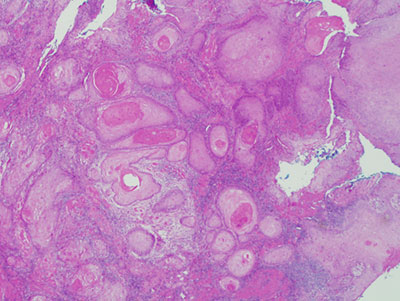
Figure 2 Low power (x40) H & E histology demonstrating neoplastic epithelial islands of variable sizes invading the connective tissue.
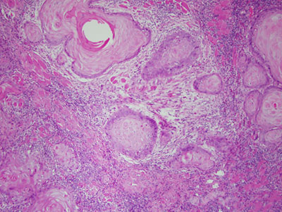
Figure 3 Higher power (x100) H & E histology closer look at the neoplastic epithelial islands sizes invading the connective tissue focusing on the cellular and nuclear morphology.
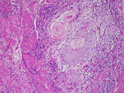
Figure 4 High power (x200) H & E histology closer ;look at the neoplastic epithelial islands invading the superficial skeletal muscle bundles.
After you have finished reviewing the available diagnostic information