Large Swelling in the Left Floor of Mouth
Can you make the correct diagnosis?
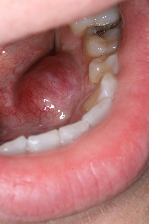
This is a 25-year-old male who presented with a mass of five years’ duration.
Sorry, you are incorrect!
The location, the prominent surface vasculature and the age of this patient are consistent with the clinical presentation of a ranula. However, the duration, the firm consistency and the pink color of the lesion are not typical of a ranula. The histology is not supportive of a ranula.
Mucoceles and ranulas are clinical terms describing exophytic, fluid-filled, fluctuant nodules, typically of minor salivary gland origin present mostly on the lower lip and the floor of the mouth. Over 90% of these lesions are cyst-like structures, or pseudocysts, and are mucous extravasation phenomena referred to as mucoceles. Some of these lesions are true cystic structures lined by epithelium and filled with mucus and are called mucus retention cysts or salivary duct cysts. The latter constitute a small percentage of all mucoceles. Ranulas, mucoceles of the floor of the mouth, constitute the other 5% and are divided into those above the mylohyoid muscle (majority) and below the mylohyoid muscle (also known as plunging ranulas or cervical ranulas). Ranulas are of minor or major salivary gland origin and are mostly extravasation in type. The etiology of the extravasation mucoceles is usually sharp trauma cutting through the salivary gland duct and releasing the mucous in the extracellular tissue. Histologically, the extravasation-type mucocele consists of a cyst-like structure lined by granulation tissue and filled with mucoid material, foamy macrophages, and at times small clusters of neutrophils. The mucous retention cysts develop as a result of a duct blockage which can be caused by trauma, fibrosis, sialolith, or pressure from an overlying tumor. The extravasation mucoceles most commonly occur on the lower lip and very rarely on the upper lip. They may occur anywhere else in the oral cavity, including the buccal mucosa and floor of mouth (Ranula). The latter can be of minor salivary gland or submandibular or sublingual gland duct origin and is more commonly seen in children and adolescents. It presents as a swelling with a bluish color if superficial, while deep mucoceles tend to take the color of the surrounding mucosa. Mucoceles tend to fluctuate in size. They are usually associated with a history of sharp lip or cheek biting, but can also be secondary to surgery in the area. This is especially true with the anterior tongue mucoceles. Surgical excision with the associated minor salivary gland is the preferred treatment for the deep mucoceles; superficial mucoceles can self-heal within 2-3 weeks. Superficial mucoceles can also mimic vesiculobullous-type diseases because they look like vesicles, especially when presenting in multiples (rare, but described). They can recur if the source of trauma is not eliminated or if they are secondary to surgery. Simple (non-plunging) ranula is best treated by marsupialization into the floor of mouth. Plunging ranula requires complete excision via an extra-oral approach. The technical difficulties associated with the complete removal of this thin-walled lesion result in a relatively high recurrence rate.
Sorry, you are incorrect!
The location, the age of the patient, the color, the firm consistency, and the duration of this lesion are all consistent with a dermoid cyst of the floor of mouth. The histology, however, is not consistent with a dermoid cyst.
Dermoid cysts of the oral cavity are rare and constitute around 1.6% of all dermoid cysts according to the original 1937 report by New and Erich. They are more common in the testis than in any other location, followed by the ovaries and the head-and-neck area. In the latter area, the floor of mouth is one of the more common areas of occurrence. This cyst is clinically classified into three types. The classification is based on its relationship to the floor of mouth muscle, the geniohyoid and mylohyoid muscles. The more common presentation is above the geniohyoid and mylohyoid muscles, which are clinically visible in the floor of mouth as they push the tongue upward, leading to dysphagia, dyspnea and dysphonia. If the cyst is between the geniohyoid and the mylohyoid muscle or below the mylohyoid muscle, it creates the appearance of a double chin. The third type is where the cyst is displaced laterally into the submandibular area. Dermoid cysts of floor of the mouth are rarely described in children under the age of 10. The majority of cases occur in patients between the ages of 10 and 30 for the floor-of-mouth cases and 15 and 40 for cases in the ovaries. Dermoid cysts above the geniohyoid muscle present as slowly enlarging large, round, raised and smooth-surfaced nodules. The nodules are usually painless unless infected, when pain can set in. Infected cysts can drain through either intraoral or extraoral fistulas. The size of the lesion determines its interference with eating, speaking and swallowing. This cyst is classified histologically into two types: cystic structures with a lumen filled with keratin and a connective tissue wall with skin adnexa and true cystic teratomas with all three germ layers tissues such as the brain, bone, muscle, respiratory and gastrointestinal tissues. The oral dermoid cysts tend to be simple with skin adnexa in the wall. The true teratoma-type cysts, which occur more often in the ovary, are prone to malignant transformation more so than those in the floor of mouth. The treatment of choice is surgical removal via intra- or extra-oral approach, depending on where the cyst is located.
Congratulations, you are correct!
Benign soft tissue neoplasms can be long-standing and can occur in the floor of mouth. For these reasons, schwannoma should be considered on the differential diagnosis. The age of the patient, the firm consistency, and the presentation as a well-circumscribed exophytic nodule are all indicative of a schwannoma. The histology is that of ancient schwannoma, a variant of this condition.
Schwannoma, also known as neurilemmoma, is a benign neoplasm of peripheral nerve origin. It is a benign, firm, well-circumscribed, smooth-surfaced, encapsulated and mobile neoplasm of Schwann cell origin. It occurs at any age but is more common in individuals between 30-50 years of age with equal sex distribution. The tongue is the most common location, followed by the floor of mouth and the lips. It is also described as occurring within the jaw bones, especially the mandible. Up to 40% of schwannomas occur in the head-and-neck area. They are usually isolated lesions unless they present as part of neurofibromatosis syndrome (type 1). Shwannoma presents as a slow-growing, firm, rubbery, smooth-surfaced nodule. Five histologic variants are described: the common/conventional type, epithelioid, plexiform, cellular, and ancient schwannoma.
Ancient schwannoma was first described by Ackerman in 1951 in the thorax and by Eversole in 1971 in the oral cavity. The clinical presentation of ancient schwannoma is not different from the common schwannoma described above. They occur in adults, with an average age of 43, and appear as well-circumscribed exophytic and smooth surfaced nodules. They can be present for up to 18 years. The size varies from 1 to 5 cm in diameter. The consistency varies from firm, as in the common type, to cystic, depending on the level of cystic degeneration affecting the lesion. Ancient schwannoma, as mentioned above, has been described in the oral cavity but is rare. These cases constitute fewer than 1% of the benign soft-tissue neoplasms of the head-and-neck area.
The histology of the common/conventional schwannoma is usually characteristic where the lesion is encapsulated and has two patterns: Antoni A, the cross section of which gives rise to “Verocay bodies,” and Antoni B, which is loose and resembles neurofibroma. Ancient schwannoma is similar to the common type in being encapsulated and with regard to the presence of Antoni A and Antoni B patterns plus Verocay bodies. It is different, however, in that it demonstrates hyalinization, cystic degeneration, focal areas of hemorrhage with hemosiderin pigment, and macrophages and hypocellularity—histologic features present in this case. Simple excision is the treatment of choice and recurrence is rare.
Sorry, you are incorrect!
There are two strong reasons to include a lipoma on the differential diagnosis: the slow-growing long duration of the lesion and the prominent surface vasculature. Otherwise, the location is rare for a lipoma, the patient is young, the color is too pink for a lipoma and the firm consistency is not that of a lipoma. The histology is not supportive of a diagnosis of lipoma.
Although rare in the mouth, lipoma has been described in the floor of mouth. Lipomas are also rare in young people and children. Lipomas are benign neoplasms of adipose tissue origin. They are more commonly described in the trunk and extremities and are rare in the oral cavity. Their overall incidence in the oral cavity constitutes around 4.4% of all benign oral lesions. In a study from the Armed Forces Institute of Pathology (AFIP) of 125 benign lipomas in and around the mouth, the male to female ratio was approximately 3:1, which is not surprising, given the study was extracted from a military population. Other studies reveal an equal gender distribution. The AFIP study showed the mean age to be 52 with a range of 9 to 92 years. Only 4 of the 125 cases involved patients under 18. In the mouth, the most common location for this neoplasm is the buccal mucosa followed by the lips, submandibular area, tongue, palate and less often on the floor of mouth and vestibule. These findings are consistent with many other published reports. These lesions are slow growing and can be present for many years. Lipomatous nodule of the buccal mucosa may represent herniation of the buccal fat pad. Lipomas usually present as a single, smooth surfaced, soft (with doughy consistency), lobulated, painless, yellowish, sessile nodule. The overlying mucosa is usually thin and stretched with visible blood vessels. Because of its softness it can be mistaken for a cyst. Histologically, lipomas are variable benign histologies. Some have predominant lobules of mature adipocytes surrounded by a thin connective tissue capsule, while others have a predominant spindle-cell component or myxoid, chondroid, connective tissue component. Some are intramuscular. Each has its own clinical behavior. Simple surgical excision is the treatment of choice for the simple, mature adipocyte component.
Sorry, you are incorrect!
A slow-growing, smooth surface firm nodule of five years’ duration can indicate a salivary gland neoplasm such as pleomorphic adenoma, which is the most common salivary gland neoplasm. However, the floor of mouth is a very unusual location for mixed tumor and the age and gender do not support that diagnosis. The histology was not consistent with pleomorphic adenoma.
Pleomorphic adenoma is the most common benign salivary gland neoplasm of both the major and minor salivary glands. It originates from the myoepithelial cells and the reserve cells of the intercalated ducts. It accounts for 80% of all benign salivary gland neoplasms. It occurs in both major and minor salivary glands and accounts for up to 77% of parotid, 68% of submandibular, and 43% of minor salivary gland tumors. It is more common in females 30-50 years of age, but it is also rarely described in children. It presents as a small, painless, slowly enlarging nodule. If left untreated it can enlarge significantly, sometimes growing to several pounds in weight. It occurs in the oral cavity, especially the palate and lips. The mixed tumor of the hard palate is fixed due to the bone-bound anatomy of the region. The tumor is otherwise movable. Histologically, mixed tumor has a wide variety of cellular and pattern manifestations; the main cellular components are epithelial duct-like structures and mesenchymal-like tissue such as myxochondroid matrix. These lesions are generally encapsulated, ranging from predominantly myxoid (36%) to extremely cellular (12%). Complete surgical removal with clean margins is the preferred treatment. Palatal lesions respond well to excision in one piece with the periosteum and overlying mucosa. The prognosis is good, but it has a tendency for recurrence (up to 44%) if not treated thoroughly. The risk of recurrence is lower in the minor salivary glands (up to 20%). The risk of malignant transformation is about 5%. The site in this case is not supportive of mixed tumor, nor is the histology.