Return to Case of the Month Archives
Enlarging mandible, spaced teeth, large tongue and coarse facial appearance
Dolphine Oda, BDS, MSc
doda@u.washington.edu
Contributed by
Dr. Ronald Snyder
Richland, WA
Case Summary and Diagnostic Information
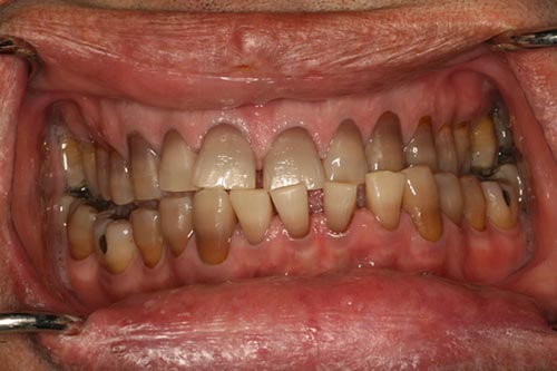
This is a 44-year-old white male who presented to Dr. Snyder as a new patient. His main complaint was that he had an under bite and that his “lower front teeth seem to be drifting and moving anteriorly” (Figure 1). He had a mountain bike accident the year before. His brother is a physician who lives in a different city had recently commented to his brother that his face “seems different.” On examination, he had a bilateral class III bite with cross bite on the left side and edge to edge on the right side. His tongue was very large and his lips (especially the lower lip) were full and had frontal bossing (Figure 2). The panoramic radiograph was not large enough to show his entire mandible A-P and left to right. His lower teeth were spaced and all of his teeth were internally dark purple in color. On further questioning, the patient also stated that his hands and feet were growing and that his gloves “keep getting too small.”
Diagnostic Information Available
This is a 44-year-old white male who presented to Dr. Snyder as a new patient. His main complaint was that he had an under bite and that his “lower front teeth seem to be drifting and moving anteriorly” (Figure 1). He had a mountain bike accident the year before. His brother is a physician who lives in a different city had recently commented to his brother that his face “seems different.” On examination, he had a bilateral class III bite with cross bite on the left side and edge to edge on the right side. His tongue was very large and his lips (especially the lower lip) were full and had frontal bossing (Figure 2). The panoramic radiograph was not large enough to show his entire mandible A-P and left to right. His lower teeth were spaced and all of his teeth were internally dark purple in color. On further questioning, the patient also stated that his hands and feet were growing and that his gloves “keep getting too small.”
Figure 1 This photograph was taken at the patient’s first clinical presentation to the dentist. Note the spacing of the mandibular teeth. The brown-gray color of the teeth is not related to this condition.
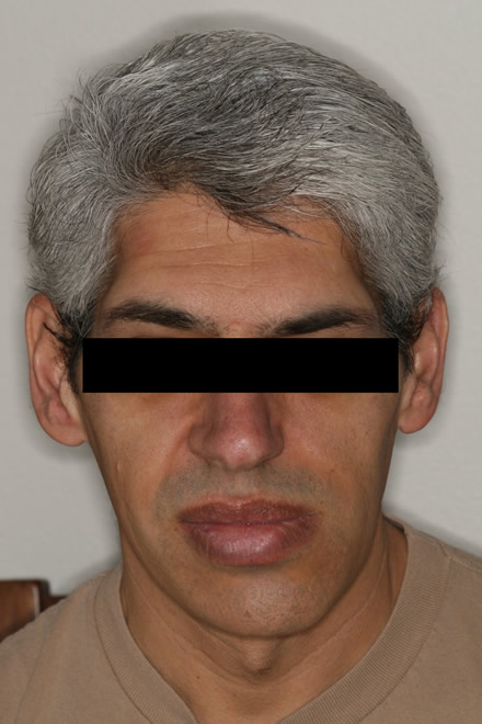
Figure 2 This photograph was taken at the patient’s first clinical presentation to the dentist. It demonstrates frontal bossing and full lips.
The past medical history is significant for asthma and hernia.
Clinical examination revealed an enlarging mandible with the appearance of prognathism (Figure 3) with spacing of teeth (Figure 2) that was not present in earlier clinical visits. His tongue appeared to have grown larger (Figure 4). Outside the mouth, he had coarse facial features which manifested as frontal bossing, full lips and a change in eye position. Upon further examination, his hands were spade-like (Figure 5) and the patient admitted that they were larger than before. Radiographically, his jaw could not be fit into the panoramic radiograph (Figure 6) because of the large size. The bony trabeculae of the mandible appeared normal but spaced out (Figure 6). The patient was referred to his primary care physician. An MRI scan of the brain showed a pituitary swelling which was biopsied.
Figure 1 This photograph was taken at the patient’s first clinical presentation to the dentist. Note the spacing of the mandibular teeth. The brown-gray color of the teeth is not related to this condition.
Figure 2 This photograph was taken at the patient’s first clinical presentation to the dentist. It demonstrates frontal bossing and full lips.
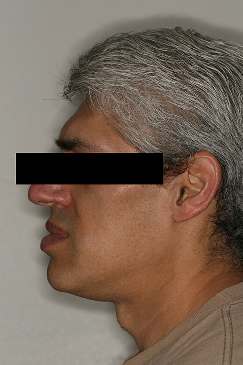
Figure 3 This photograph was taken at the patient’s first clinical presentation to the dentist. It demonstrates mandibular prognathism.
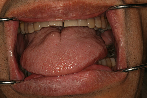
Figure 4 This photograph was taken at the patient’s first clinical presentation to the dentist. It demonstrates a large tongue
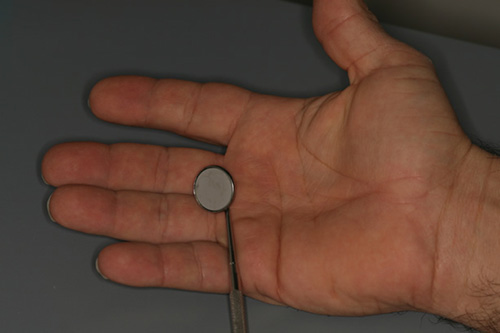
Figure 5 This photograph was taken at the patient’s first clinical presentation to the dentist. It demonstrates large, spade-like hands.
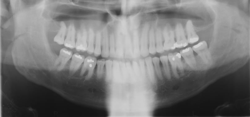
Figure 6 This panoramic radiograph demonstrates a large mandible. The entire mandible could not be fit in the panoramic radiograph.
Treatment
Under general anesthesia, the patient underwent surgery for the pituitary tumor. Further imaging of the pituitary gland subsequent to surgery revealed residual tumor. He underwent radiation therapy for the residual tumor and has been disease-free since.
Incisional and excisional biopsy
Histological examination of the pituitary tumor revealed sheets of uniform cells, many with granular eosinophilic cytoplasm with central nuclei and some exhibiting more chromophobic cytoplasm and round-to-oval nuclei eccentrically located (Figure 7). Prominent nucleoli were present as well as cells with multiple nuclei. Immunohistochemistry staining revealed uniform and diffuse positive staining with antibody to growth hormone (Figure 8) and focal (minority cells) positive staining with antibody to prolactin (Figure 9).
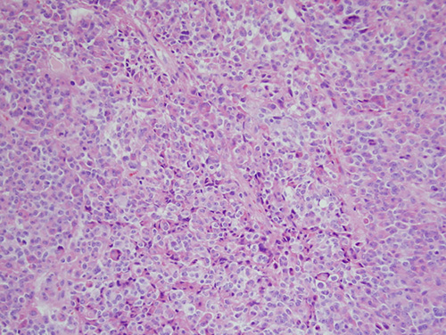
Figure 7 Low power (x100) H & E histology reveals sheets of uniform cells, many with granular eosinophilic cytoplasm with central nuclei and some with more chromophobic cytoplasm and round-to-oval nuclei eccentrically located. Prominent nucleoli were present as well as cells with multiple nuclei.
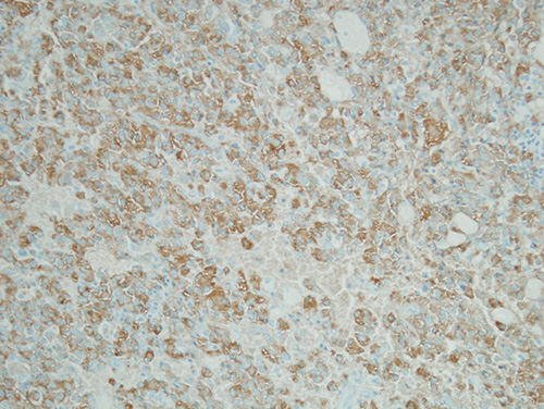
Figure 8 High power (x200) immunohistochemistry stain reveals uniform and diffuse positive staining with antibody to growth hormone.
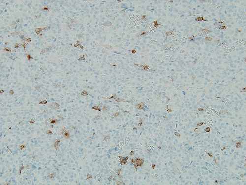
Figure 9 High power (x200) immunohistochemistry stain reveals focal (minority cells) positive staining with antibody to prolactin (Figure 9).
After you have finished reviewing the available diagnostic information