Partially Corticated, Expansile & Destructive Radiolucency Between Teeth #s 28 & 30
Can you make the correct diagnosis?
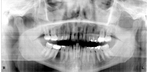
This is a 53-year-old Asian female who presented with an expansile and well-demarcated radiolucency involving teeth #s 28 & 30 of 18 months’ duration.
Sorry! you are incorrect
The bony expansion, the location and the bony perforation are all typical clinical presentations for ameloblastomas in general, which could include a diagnosis of unicystic ameloblastoma. The age of the patient, however, is advanced compared to that in most cases of unicystic ameloblastoma. Furthermore, 90% of unicystic ameloblastomas present with impacted teeth simulating a dentigerous cyst, whereas the patient in this case does not have any impacted teeth. The histology in this case was not supportive of unicystic ameloblastoma.
Ameloblastomas are a family of neoplasms with varied clinical and radiographic presentation. Among these neoplasms, unicystic ameloblastoma would be the more likely diagnosis in this case if the unilocular radiolucency were taken into account as the most significant finding. However, neither the age of the patient nor the perforation of the jaw bone is supportive of a diagnosis of unicystic ameloblastoma. On the other hand, the age and bony perforation is a common presentation for the conventional (solid, multicystic) ameloblastoma. The latter, however, tends to be radiographically multilocular rather than unilocular. In addition, 90% of unicystic ameloblastomas occur in association with an impacted tooth resembling a dentigerous cyst. As noted, the histology in this case was not supportive of any type of ameloblastoma. The patients with unicystic ameloblastomas are typically much younger, with an age range of 14-20 years.
Ameloblastoma, if not treated, can reach very large sizes, causing facial disfigurement. It loosens, displaces and resorbs adjacent teeth. Ameloblastomas are usually not painful unless infected, in which case they can be mildly painful. Parasthesia and anesthesia are extremely rare, unless the lesion is very large in size. Also, ameloblastoma tends to expand rather than perforate the cortical bone; if the latter occurs with extension into the adjacent soft tissue, it has a higher tendency for recurrence and therefore would have a worse prognosis than cases in which the ameloblastoma is completely encased by bone. The solid type is treated with en bloc or resection with clean margins. Curettage is the treatment of choice for the unicystic ameloblastoma.
Congratulations! You are correct
The age of the patient, the location, and the apparent perforation of the bone all support a diagnosis of odontogenic keratocyst (OKC). For that reason, OKC should be placed high on the differential diagnosis list. However, the described expansion of the bone is unlike the typical behavior of an OKC, which hollows bone rather than causing it to expand. Nonetheless, the histology is supportive of an OKC.
Odontogenic keratocyst is an aggressive cyst known for its rapid growth and its tendency to invade the adjacent tissues, including bone. It has a high recurrence rate and is associated with bifid rib basal cell nevus syndrome. The majority of patients are in the age ranges of 20-29 and 40-59, but cases in patients ranging in age from 5 to 80 years have been reported. The distribution between sexes varies from equal distribution to a male-to-female ratio of 1.6:1, except in children. Odontogenic keratocyst predominantly affects Caucasian populations and, if one may judge from the limited evidence provided by the literature, is chiefly of Northern European descent.
Odontogenic keratocysts may occur in any part of the upper and lower jaw, with the majority (almost 70%) occurring in the mandible. They occur most commonly in the angle of the mandible and ramus. Posterior mandible is an area common to many benign odontogenic tumors such as ameloblastoma and odontogenic myxoma and is also a typical location for dentigerous cysts. Radiographically, OKCs present predominantly as unilocular radiolucencies with well-defined, sclerotic or scalloped borders. They may also present as multilocular radiolucencies. Odontogenic keratocysts of the maxilla are smaller in size when compared to those occurring in the mandible; larger OKCs tend to expand bone, but mildly—obvious clinical expansion (which is the case in this patient) should be viewed with suspicion for a neoplasm. OKCs can also present as small and oval radiolucencies between teeth simulating a lateral periodontal cyst, in an area of an extracted tooth simulating a residual cyst, at the apex of a vital tooth mistaken for a periapical cyst, or in the anterior maxilla between the central incisors simulating an incisive canal cyst. OKCs grow to sizes larger than any other odontogenic cysts. They usually penetrate the bone rather than expand it and grow in an anterior to posterior direction. Despite this aggressive growth, they often remain asymptomatic, thus growing to large sizes and hollowing the bone.
Odontogenic keratocysts are significant clinical entities due to their tendency for recurrence and destructive behavior. They are known to have a high recurrence rate, ranging from 13% to 60% (1, 2). Complete surgical removal is the treatment of choice. Surgery includes enucleation, curettage, enucleation with peripheral ostectomy, and resection depending on the radiographic presentation, location and clinical behavior. Surgery combined with Carnoy’s solution or liquid nitrogen treatment has been effective in reducing the recurrence rate. At times, adjacent or associated teeth are extracted in the interest of complete removal. Some investigators advocate marsupialization and occasionally resection of the more aggressive cysts that tend to perforate buccal and lingual bone. Resection is a rare modality of treatment. Most cysts recur within the first three years while others may recur as late as after 16 years. Conservative surgical removal and long-term follow-up is the treatment of choice by most clinicians.
Sorry! you are incorrect
The described bone expansion and perforation of bone are typical of the behavior of a neoplasm, including a neoplasm of tooth origin such as an odontogenic myxoma. The age of the patient, however, is not consistent given that odontogenic myxomas rarely occur after 50 years of age. Nonetheless, odontogenic myxoma should be placed high on the differential diagnosis. The histology of this case is not supportive of an odontogenic myxoma.
Odontogenic myxoma occurs in the jaw bones, usually in the tooth-bearing areas of the jaw. It is an uncommon, benign, but locally aggressive neoplasm. Nearly all cases so far have been described in the jaw bones. Therefore, it is of tooth origin, and is believed to be from the mesenchymal portion of a tooth germ, most likely of the dental papilla. It has the potential for extensive bony destruction and extension into the surrounding structures. It is less common than odontomas and ameloblastomas. For that reason, a pathologist who is not familiar with the histology of a tooth germ can mistake a myxoid dental follicle for an odontogenic myxoma. Almost 75% of odontogenic myxomas occur in patients around 23-30 years of age with a slight female predilection (1:1.5 male-to-female). It rarely occurs in patients over 50 or under 10 years of age. It occurs almost equally in the maxilla and mandible with a slight predilection for the posterior mandible. A few cases are described in the ramus and condyle, non-tooth bearing areas.
Odontogenic myxoma is slow growing, persistent and destructive. Most cases are expansile and can displace and resorb teeth. In the maxilla, they usually invade the maxillary sinuses and at times (though rarely) cross the midline to the opposing sinus. Radiographically, the majority present as expansile and multilocular, though some are unilocular with or without scalloped borders, and rare cases present with a diffuse and mottled appearance which can be mistaken for a malignant neoplasm. Grossly, this lesion is gelatinous in nature, making curettage alone difficult; the more fibrotic odontogenic myxomas (also known as odontogenic myxofibroma or fibromyxoma) have more body and are easier to curette. Histologically, it is made up of loose and delicate fibrous connective tissue. The fibroblasts are stellate and are suspended on a delicate network of collagen fibrils. Immunohistochemistry studies suggest that the spindle-shaped cells constituting this neoplasm have a combined fibroblastic and smooth muscle typing, suggesting that it is of myofibroblastic origin. Small blood vessels are present, as are small odontogenic epithelial islands on occasion. Sometimes, this lesion is fibrotic, making it easier to curette. The treatment of choice is surgical excision ranging from segmental resection with clear bony margins of up 1.5cm to prevent recurrence of the neoplasm. Curettage with and without cauterization is used for treatment, but is associated with a high recurrence rate. Reconstruction can be immediate or delayed, and can include an autologous bone graft from the anterior or posterior iliac crest. Fibula-free vascular osteocutaneous bone graft is another reconstructive modality, as is distraction osteogenesis. Immediate postoperative follow up is weekly for approximately one month, then monthly for the next five months and twice a year for the next five years.
Sorry! you are incorrect
The location anterior to the first molar, the bony expansion, the radiographic presentation of radiolucency, the bony perforation and the gender of this patient are compatible with some aspects of CGCG. However, the age of the patient is not, and neither is the histology.
Jaffe first coined the term “reparative” for central giant cell granuloma. Most pathologists have since dropped the term “reparative” for lack of evidence that the pathogenesis is a reparative process. CGCG is described as a non-neoplastic process and yet can behave in a very aggressive and expansile manner, destroying bone and displacing teeth. Over 60% of CGCG cases occur in patients younger than 30 years of age, with twice as many occurrences in females as in males. CGCG is classified into aggressive and non-aggressive types; the aggressive type tends to occur in younger patients and causes disfiguration, especially after surgery. Over 70% of cases occur in the mandible anterior to the first molar tooth. This lesion has also been described in other cranio-facial and small long bones such as those of the hands and feet.
The usual treatment for CGCG is surgery, ranging from curettage and en bloc to resection. The latter is used in aggressive and recurring cases. In the past ten years or so, alternatives to surgery have emerged with successful results, saving some patients from facial disfigurement. Steroid injections are the most successful alternative treatment thus far; they require injections weekly or every 2-3 weeks, have no known side effects (even in children), and are the least expensive alternative treatment. Other treatments include: calcitonin injections or nasal spray, which require daily injections or a nasal spray of salmon calcitonin for about a year and are safe for pregnant females; and interferon alfa-2a injections, which are administered 2-3 times per week for several months and are the most expensive alternative treatment.