Mixed Radiolucent Radiopaque Lesion Associated with Impacted Tooth #14
Can you make the correct diagnosis?
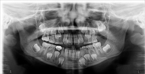
This is a 7-year-old female who first presented to the Center for Pediatric Dentistry with a chief complaint of pain associated with cracked tooth #D.
Sorry! you are incorrect
The clinical presentation of a lesion around an impacted tooth should make one think first and foremost (if not only), of tooth related (odontogenic) conditions. In this case, the lesion is radiolucent/radiopaque which is limiting the differential diagnosis to a few conditions and odontoma would be the most likely disease given the age and the location. However, the hard-tissue produced by this case is not in form of small masses simulating tooth-like structures. For that reason, odontoma would be a less likely diagnosis. Nonetheless, odontoma stays on the differential diagnosis. The histology was not supportive of an odontoma.
Whether complex or compound, hamartoma or neoplasm, odontoma is the most common odontogenic tumor, accounting for 22% of all odontogenic tumors. It is of mixed epithelial and mesenchymal origins. It is usually associated with unerupted teeth. It can occur at any age, but is most common in the first two decades of life, with an average age of 14 or 18. It is slightly more common in females and more common in the maxilla, especially the anterior maxilla, than in the mandible. Compound odontomas are more common in the anterior jaws, while complex odontomas occur more often in the posterior jaws. In about 80% of cases, they are associated with impacted or unerupted teeth. Radiographically, odontomas present as a well-circumscribed radiolucency resembling a dental follicle or dentigerous cyst with one or multiple radiopaque pieces resembling teeth. Compound odontomas tend to occur between teeth and tend to be composed of multiple small tooth-like structures, while complex odontomas tend to occur in the posterior jaws and present as a conglomerate mass. Both types are made up of enamel matrix, dentin, cementum, and dental pulp surrounded by a dental follicle or cyst. Histologically, the tooth-like structures are arranged in a uniform manner similar to the normal tooth. The structures in complex odontomas are mixed and disorganized. These lesions are benign and are conservatively treated with simple curettage. Recurrence is not described; if it recurs, one must rule out other odontogenic lesions such as calcifying odontogenic cyst and ameloblastic fibro-odontoma.
Congratulations! You are correct
With the exception of the location being in the maxilla, everything else fits well with the clinical and radiographic presentation of ameloblastic fibro-odontoma.
Ameloblastic fibroma-odontoma is a relatively uncommon neoplasm. It is a benign, slow-growing, expansile, neoplasm or hamartoma of mixed epithelial and mesenchymal odontogenic origins. Some suggest that this lesion may represent an early stage of an odontoma. However, it behaves like a neoplasm in terms of growth and expansion of the jaw bones. It is known to inhibit tooth eruption and is therefore often associated with impacted teeth. It can also displace teeth. Therefore, these lesions can go undetected by parents unless a noticeable expansion occurs or because of failure of tooth eruption and displacement of teeth. It is rarely painful. It occurs most commonly in children in the first and second decade of life with an age range of infancy to 42 years of age. It has equal gender predilection and is more common in the posterior mandible than in the maxilla. It also occurs more often in the posterior jaws than in the anterior jaws. Radiographic findings include an impacted/unerupted tooth with a well-defined, usually unilocular radiolucency around the crown of a tooth, similar to a dentigerous cyst. It may also present as a radiolucency containing radiopaque material ranging from flecks to small tooth-like structures. Because of the continued slow growth, this lesion can reach very large sizes if left untreated. The histology of ameloblastic fibro-odontoma is made up of mixed epithelial and mesenchymal components. The epithelial component presents in the form of small islands, cords and rosettes of epithelial cells with columnar and palisaded peripheries and with centers that may or may not contain stellate or cuboidal epithelial cells. The mesenchymal component is made up of primitive connective tissue stroma with tooth-like hard material that has the features of a complex odontoma. They are benign and well-differentiated neoplasms; for that reason, transformation is rarely described. Conservative curettage and enucleation is the treatment of choice, and there is a very low recurrence rate. The involved tooth is usually extracted. It has a good prognosis.
References
- Vengal M, Arora H, Ghosh S. Large erupting complex odontoma: a case report. J Can Dent Assoc. 2007 Mar;73(2):169-73.
- Noonan VL, Gallagher G, Kabani S. Compound and complex odontomas. J Mass Dent Soc. 2006 Fall;55(3):40.
- Ogunlewe MO, Adeyemo WL, Ladeinde AL. Surgical management of a large complex odontoma of the mandibular angle-ramus region through intra-oral buccal approach–A case report. Niger Postgrad Med J. 2005 Dec;12(4):312-5.
- Chen Y, Li TJ, Gao Y. Ameloblastic fibroma and related lesions: a clinicopathologic study with reference to their nature and interrelationship. J Oral Pathol Med. 2005 Nov;34(10):588-95.
- Dhanuthai K, Kongin K. Ameloblastic fibro-odontoma: a case report. J Clin Pediatr Dent. 2004 Fall;29(1):75-7.
- Oghli AA, Scuto I, Ziegler C. A large ameloblastic fibro-odontoma of the right mandible. Med Oral Patol Oral Cir Bucal. 2007 Jan 1;12(1):E34-7.
- Praetorius F, Hjorting-Hansen E, Gorlin RJ, Vickers RA. Calcifying odontogenic cyst: range, variations and neoplastic potential. Acta Odonotol Scand 1981;39:227-240.
- Gorlin RJ, Pindborg JJ, Clausen FP, Vickers RA. The calcifying odontogenic cyst: a possible analogue of the cutaneous calcifying epithelioma of Malherbe. Oral Surg Oral Med Oral Pathol 1962;15:1235-1243.
- Hong SP, Ellis GL, Hartman KS. Calcifying odontogenic cyst: a review of ninety-nine cases with reevaluation of their nature as cysts or neoplasms, the nature of ghost cells, and subclassification. Oral Surg Oral Med Oral Pathol 1991;72:56-64.
- Batra P, Prasad S et al. Adenomatoid odontogenic tumour: review and case report. J Can Dent Assoc. 2005; 71:250-253.
- Philipsen HP, Birn H. The adenomatoid odontogenic tumor, ameloblastic adenomatoid tumor or adeno-ameloblastoma. Acta Pathol Microbiol Scand 1969; 75:375–398.
- Philipsen HP, Reichart PA, Zhang KH, Nikai H, Yu QX. Adenomatoid odontogenic tumor: biologic profile based on 499 cases. J Oral Pathol Med 1991; 20:149–158.
- Sato D, Matsuzaka K et al. Adenomatoid odontogenic tumor arising from the mandibular molar region: a case report and review of the literature. Bull Tokyo Dent Coll. 2004; 45:223-227.
- Ungari C, Poladas G, Giovannetti F, Carnevale C, Iannetti G. Pindborg tumor in children. J Craniofac Surg. 2006 Mar;17(2):365-9.
- Guerrisi M, Piloni MJ, Keszler A. Odontogenic tumors in children and adolescents. A 15-year retrospective study in Argentina. Med Oral Patol Oral Cir Bucal. 2007 May 1;12(3):E180-5.
Sorry! you are incorrect
Another hard-tissue forming condition of odontogenic origin that should be included on the differential diagnosis is calcifying odontogenic cyst (COC). The location is acceptable but the patient’s age is on the very young end of the spectrum. The histology, however, is not supportive of this diagnosis.
Calcifying odontogenic cyst (COC) constitutes around 1% of all odontogenic cysts. Praetorius et al were the first to classify COC into two types: a simple cyst and a solid neoplastic spectrum. They classified the cystic type into subtypes A, B and C: A denoting a simple cyst, B an odontoma-producing cyst, and C a cyst with ameloblastomatous proliferating. COC tends to occur around the third decade, with a patient age range of 7-82 years. It occurs equally in the maxilla and mandible, usually anterior to the first permanent molar, though it has a predilection for occurrence in the maxilla in the younger age range. It occurs equally in males and females. COC occurs more commonly in bone, but it can also occur in soft tissue (in the gingiva without a bony component, also known as peripheral COC). The peripheral counterpart of both lesions is less aggressive than the intra-osseous type. Histologically, COC can present in several ways: as a simple cyst with ghost cells and focal ameloblastoma-like epithelial changes, with more proliferative epithelium and ghost cells, with calcifications (both amorphous, tooth-like and calcified ghost cells), or as a true odontoma associated with such a cyst. Radiographically, COCs tend to be well-defined radiolucencies with occasional small radiopacities. They can be present at the apex, between teeth, or associated with impacted teeth. Clinically, COC can both expand the jaws and extend into the surrounding soft tissues and even the maxillary sinus. It can also resorb and displace teeth. The treatment of choice is thorough curettage.
Sorry! you are incorrect
The patient’s age and the location are not supportive of Pindborg tumor. The radiolucent/radiopaque presentation associated with an impacted tooth is primarily the reason why Pindborg tumor is on the differential diagnosis. The histology is not supportive of Pindborg tumor.
Calcifying epithelial odontogenic tumor (CEOT, Pindborg tumor) was first described in 1956. It is a rare odontogenic neoplasm which constitutes less than 1% of all odontogenic tumors. It is believed to originate from the stratum intermedium. Pindborg tumor is described at a wide age spectrum but is clustered around 30-50 years of age, with equal gender distribution. It is therefore rare in children. It is also more common in the posterior mandible; around 75% of cases occur in this area. It is expansile but otherwise asymptomatic. Slightly more than 50% of instances of this lesion are associated with impacted teeth. Peripheral or extra-osseous Pindborg tumor has been described mainly on the gingiva, more so on the anterior gingiva. One peripheral Pindborg has been described on the upper lip. Radiographically, CEOT presents in several patterns ranging from well-circumscribed unilocular radiolucency with radiopacity to multilocular, “honeycomb” radiolucency with flecks of radiopaque material. The pattern of “driven snow” has been attached to this lesion. It is histologically made up of sheets, islands and small cords of polyhedral epithelial cells with some evidence of pleomorphism. However, it is a benign neoplasm and therefore pleomorphism should not alter the diagnosis. Because of the latter, this lesion is frequently misdiagnosed as a metastatic disease. Intercellular bridges are prominent. These cells produce and release amyloid, an unusual substance for a tooth-related neoplasm to be producing, but nonetheless, it does. Resection or en bloc is the treatment of choice. This may result in mandibular discontinuity requiring secondary reconstruction. The overall prognosis for Pindborg tumor is good.