October 2009: Gingival swelling left posterior mandible
Can you make the correct diagnosis?
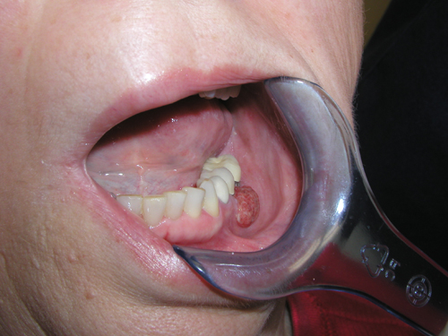
This is a 65-year-old white female with a rapidly enlarging gingival swelling interproximal to teeth #s 19 & 20. This swelling was of three weeks’ duration and is red and ulcerated. The lesion was not painful and the patient’s only complaint was that the lesion was growing larger. It was 0.8 x 1.2 cm in greatest dimensions. Radiographs were negative for any bony changes. The patient denied tobacco use. She drinks 1-2 glasses of wine per week.
Sorry! you are incorrect
One of the most common gingival swellings is pyogenic granuloma; for that reason, it should be high on the differential diagnosis list. PG can grow fast and is usually exophytic and red consistent with the clinical presentation of this case. The histology, however, is not consistent with PG.
Pyogenic granuloma constitutes 85% of all reactive gingival swellings, representing a profuse mass of vascular granulation tissue (1). It can be induced by local irritants such as excessive plaque, sharp fillings and dental calculus; it sometimes forms in an extraction socket in response to an irritant left in the socket. It can occur anywhere in the oral cavity and skin, especially the tongue, lips, fingers and nail beds (1). In the mouth, it occurs most commonly in the gingiva, especially the maxillary buccal and interproximal gingiva (1-2). Occasionally, it may surround the tooth. It is usually highly vascular, fast-growing, exophytic, lobular, sessile, and ulcerated or covered by pseudomembrane. The color changes from red to pink when it starts to heal. It occurs at any age and sex with a slight predilection for young females; it affects 1% of pregnant females. Pyogenic granuloma is usually painless except during eating, when bleeding and pain is described (1). Histologically, it presents as a mass of loose and vascular granulation tissue, usually with ulcerated or eroded surface epithelium and many inflammatory cells. A range of treatment modalities are available, including excision with removal of the local irritant, laser surgery, or intralesional injection with absolute alcohol, steroids or botulinum toxin (2-3). Scaling and polishing prior to surgical removal helps shrink the lesion. The prognosis is good, although recurrence is possible, especially during pregnancy.
Sorry! you are incorrect
The third most common gingival swelling is PGCG. The exophytic clinical presentation can be supportive of PGCG but not so much the color. PGCG tends to be more grayish blue in color and this lesion is bright red. PGCG is an exclusively gingival swelling and can grow fast. The histology, however, is not consistent with PGCG.
Peripheral giant cell granuloma constitutes fewer than 5% of all reactive gingival swellings, and consists of a hyperplastic mass of vascular granulation tissue with many osteoclast-like multinucleated giant cells. It presents as a lobular, purplish-blue exophytic nodule exclusively on the gingiva, both edentulous and dentate, and usually anterior to the molars (4-6). It originates from the periodontal ligament or the periosteum. It occurs across a wide age range, commonly in children, young adults, and in females (2:1 female to male ratio) (6). It presents as sessile or pedunculated and smooth surfaced or lobular, and though usually painless it can occasionally be ulcerated, painful and accompanied by bleeding (4-6). Like pyogenic granuloma, it is usually present either on the buccal or lingual gingiva or between teeth, but it can occasionally surround the teeth (5) and act aggressively by displacing teeth much like a sarcoma (4). It can also resorb the underlying bone in a smooth and concave “saucer-like” manner. Complete excision, including curettage of underlying bone, is the preferred treatment. It has a good prognosis with a recurrence rate of approximately 10% (4-6).
Sorry! you are incorrect
The second most common gingival swelling is peripheral ossifying fibroma and therefore should be considered on the differential diagnosis. The bright red color however is not supportive of POF, neither is the histology.
Peripheral ossifying fibroma constitutes 10% of all reactive gingival swellings. It consists of a moderately cellular fibrous connective tissue mass with bony trabeculae and/or cementum-like hard tissue. It has been reported rarely on edentulous alveolar mucosa. It originates from the periodontal ligament or the periosteum. This lesion is most common in young patients between 1 and 19 years of age and has a predilection for females over males by a 3:2 ratio (7-9). It occurs exclusively on the gingiva, especially the anterior gingiva, with slight predilection to the maxilla and rare presentation in primary teeth (9). It is usually sessile and exophytic and often ulcerated; it presents as well-demarcated sessile nodules, which are firm or hard depending on the amount of ossification and calcifications (7-8). Peripheral ossifying fibroma is usually pink but can be focally red if ulcerated. Histologically, peripheral ossifying fibroma is made up of a moderately cellular mass of fibrous connective tissue with calcifications ranging from cementum-like material to calcified bony trabeculae with viable osteocytes. The surface epithelium overlying the mass is usually ulcerated. Deep surgical excision to include the periodontal ligament is the preferred treatment, though laser removal has been used effectively. Deep surgery may lead to a gingival defect, which would require gingival grafting, especially if it is located on the anterior buccal gingiva (8). There is a 16-20% recurrence rate (9).
Congratulations! You are correct
Squamous cell carcinoma of the gingiva is uncommon, especially in a non-smoker and social alcohol user. Oral squamous cell carcinoma of the mouth is a highly aggressive neoplasm that currently ranks as the fifth most common malignant neoplasm worldwide and accounts for an estimated 90% of oral malignancies (10). Oral SCC occurs predominantly in males over the age of 40 years, with an observed male-to-female ratio of 2:1 generally and 1.4:1 in the USA (10-12). Excluding the outer lip, the most common sites (in decreasing order) are the ventral and lateral surfaces of the tongue (25-50%), floor of mouth (15%), gingiva (12%) and palate (9%). The buccal mucosa and retromolar pad areas (3%) have a relatively low incidence of occurrence (11) unless the patient is a chronic smokeless tobacco user. Oral SCC varies in presentation from deceptively innocent-looking to obviously malignant. It may present as a non-healing ulcer, or as red, white or mixed red-and-white lesions. Characteristic signs of oral SCC are non-healing ulcer, ulcer with rolled borders, fungation, fixation and induration. Rarely, oral SCC may present as unexplained asymptomatic lateral neck lymphadenopathy (10-12). Oral SCC is most commonly associated with chemically induced mutagenesis, specifically tobacco and alcohol use (11).
Tobacco use is described in over 75% of oral SCC patients (10-12). Tobacco and alcohol have been shown to act synergistically in the development of oral SCC. Human papilloma virus (HPV) has also been found to have a high prevalence in oral cancer, especially in younger patients with no history of tobacco use. Other factors include poor oral hygiene, syphilis, chronic candidiasis, iron and dietary deficiencies, herpes simplex and various other immunologic factors, and lichen planus—especially persistent erosive lichen planus.
Determination of the prognosis of oral SCC is based on its clinical stage and histological classification (10-12). Although oral SCC is a diagnosis made by histology, surgeons tend to depend exclusively on the TNM classification system for clinical staging and treatment decisions. Prognosis is dependent on the TNM staging system; the most important prognostic sign is the presence or absence of metastases at the time of diagnosis. The prognosis thus improves when the lesion is detected early. Oral SCC patients die mainly of infection due to lowered resistance or of hemorrhage if the tumor erodes through one of the main blood vessels.
References
- Fantasia JE, Damm DD. Red nodular lesion of tongue. Pyogenic granuloma.
Gen Dent. 2003 Mar-Apr;51(2):190-194. - Ichimiya M, Yoshikawa Y, Hamamoto Y, Muto M. Successful treatment of pyogenic granuloma with injection of absolute ethanol. J Dermatol. 2004 Apr;31(4):342-4.
- Pham J, Yin S, Morgan M, Stucker F, Nathan CA. Botulinum toxin: helpful adjunct to early resolution of laryngeal granulomas. J Laryngol Otol. 2004 Oct;118(10):781-5.
- Flaitz CM, Peripheral giant cell granuloma: a potentially aggressive lesion in children. Pediatr Dent. 2000 May-Jun;22(3):232-3.
- Chaparro-Avendano AV, Berini-Aytes L, Gay-Escoda C. Peripheral giant cell granuloma. A report of five cases and review of the literature. Med Oral Patol Oral Cir Bucal. 2005 Jan-Feb;10(1):53-7; 48-52.
- Neville BW, Damm DD, Allen CM, Bouquot JE. Peripheral giant cell granuloma. In: Oral and Maxillofacial Pathology, 2nd edition. Philadelphia: W.B. Saunders, 2002. p. 449-451.
- Hanemann JA, Pereira AA, Ribeiro Junior NV, Oliveira DT. Peripheral ossifying fibroma in a child: report of case. J Clin Pediatr Dent. 2003 Spring;27(3):283-5.
- Walters JD, Will JK, Hatfield RD, Cacchillo DA, Raabe DA. Excision and repair of the peripheral ossifying fibroma: a report of 3 cases. J Periodontol. 2001 Jul;72(7):939-44.
- Cuisia ZE, Brannon RB. Peripheral ossifying fibroma–a clinical evaluation of 134 pediatric cases. Pediatr Dent. 2001 May-Jun;23(3):245-8
- Bundgaard T, S Bentzen, et al. Histopathologic, stereologic, Epidemiologic, and clinical parameters in the prognostic evaluation of squamous cell carcinoma of the oral cavity. Head & Neck. 18:142-152 (1996).
- Barasch A, DE Morse, et al. Smoking, gender, and age as risk factors for site-specific intraoral squamous cell carcinoma. Cancer 73:509-513 (1994).
- Syrjanen SM, KJ Syrjanen et al. Human papillomavirus (HPV) DNA sequences in oral precancerous lesions and squamous cell carcinoma demonstrated by in situ hybridization. J Oral Pathol. 17:273 (1988).