Extensive Radiolucency Posterior Maxilla
Can you make the correct diagnosis?
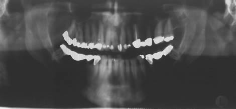
This is a 60-year-old white male who was referred to Dr. Mark Carlson an Oral and Maxillofacial Surgeon from Tacoma to evaluate a severe and localized bone loss in the upper right posterior maxilla.
Sorry! you are incorrect
Odontogenic keratocyst is an aggressive cyst known for its rapid growth and its tendency to invade the adjacent tissues, including bone. It has a high recurrence rate and is associated with bifid rib basal cell nevus syndrome (1, 2). The majority of patients are in the age ranges of 20-29 and 40-59, but cases in patients ranging in age from 5 to 80 years have been reported. The distribution between sexes varies from equal distribution to a male-to-female ratio of 1.6:1, except in children (1, 2). Odontogenic keratocyst predominantly affects Caucasian populations and, if one may judge from the limited evidence provided by the literature, is chiefly of Northern European descent (1).
Odontogenic keratocysts may occur in any part of the upper and lower jaw, with the majority (almost 70%) occurring in the mandible. They occur most commonly in the angle of the mandible and ramus (1). Posterior mandible is an area common to many benign odontogenic tumors such as ameloblastoma and is also a typical location for dentigerous cysts. Radiographically, OKCs present predominantly as unilocular radiolucencies with well-defined, sclerotic or scalloped borders. They may also present as multilocular radiolucencies. Odontogenic keratocysts of the maxilla are smaller in size when compared to those occurring in the mandible; larger OKCs tend to expand bone, but mildly—obvious clinical expansion should be viewed with suspicion for a neoplasm. OKCs can also present as small and oval radiolucencies between teeth simulating a lateral periodontal cyst, in an area of an extracted tooth simulating a residual cyst, at the apex of a vital tooth mistaken for a periapical cyst, or in the anterior maxilla between the central incisors simulating an incisive canal cyst (2). OKCs grow to sizes larger than any other odontogenic cysts. They usually penetrate the bone rather than expand it and grow in an anterior to posterior direction (3). Despite this aggressive growth, they often remain asymptomatic, thus growing to large sizes and hollowing the bone.
Odontogenic keratocysts are significant clinical entities due to their tendency for recurrence and destructive behavior. They are known to have a high recurrence rate, ranging from 13% to 60% (1, 2). Complete surgical removal is the treatment of choice. Surgery includes enucleation, curettage, enucleation and peripheral ostectomy, to resection depending on the radiographic presentation, location and clinical behavior. Surgery combined with Carnoy’s solution or liquid nitrogen treatment have been effective in reducing recurrence rate (1, 2). At times, adjacent or associated teeth are extracted in the interest of complete removal. Some investigators advocate marsupialization and occasionally resection of the more aggressive cysts that tend to perforate buccal and lingual bone. Resection is a rare modality of treatment. Most cysts recur within the first three years while others may recur as late as after 16 years (1, 2). Conservative surgical removal and long-term follow-up is the treatment of choice by most clinicians. The histology of this case is not supportive of Odontogenic keratocyst.
Congratulations! You are correct
Ameloblastoma is one of the most common benign neoplasms of odontogenic origin. It accounts for 11% of all Odontogenic neoplasms/hamartomas (4-6). It is a slow-growing, persistent, and locally aggressive neoplasm of epithelial origin. It affects a wide range of age distribution but is mostly a disease of adults, at an average age of 33, with equal sex distribution. Reports from Africa and India show male predilection. It has a prevalence to patients of black origin (5).
About 85% of ameloblastomas occur in the posterior mandible, most in the molar-ramus area, some in the anterior mandible. About 15% occur in the maxilla, the vast majority in the posterior maxilla as is the case in our patient. Ameloblastomas of the maxilla tend to occur in older age population consistent with our patient (4-6). Ameloblastomas of the maxilla tend to have a worse prognosis than those of the mandible. They tend to recur more and behave more aggressively in terms of early invasion of the maxillary sinus (4, 6). They can also invade the optical nerve and the brain and kill the patient. The notion of ameloblastomas of the maxilla being more aggressive in behavior may be biological in nature such as the histologic type (plexiform ameloblastoma reported to be more aggressive than follicular ameloblastoma) or because of the anatomic location being difficult to reach surgically making complete eradication of a the neoplasm a challenge.
Ameloblastoma is rarely described in children. It is characteristically expansile, radiolucent and multilocular in nature. It can however be unilocular and associated with impacted teeth resembling a dentigerous cyst (4-6). The latter are known to be less aggressive than the multilocular solid lesions. Three clinical types of ameloblastoma are described, the solid type (radiographically multilocular), the cystic (radiographically unilocular and usually associated with an impacted tooth) and peripheral type (soft tissue usually gingival ameloblastoma) (4-6). Our case qualifies for the solid type, although radiographically was not multilocular in appearance. For more information on the behavior of the various types of ameloblastomas, please read the October 04 newsletter under clinical case discussion.
Ameloblastoma, if not treated, can reach very large sizes with facial disfiguring. It loosens, displaces and resorbs adjacent teeth. With the exception of jaw expansion, it is usually asymptomatic unless infected where it can be mildly painful. Parasthesia, anesthesia is extremely rare, unless they are very large in size. Also, ameloblastoma tends to expand rather than perforate the cortical bone. If the later occurs with extension into the adjacent soft tissue, it would have a higher tendency for recurrence and therefore would have a worse prognosis than those completely encased by bone (4-6).
As mentioned above, three clinical types of ameloblastomas are described, the solid being the most common is further subtyped histologically into follicular (most common type), acanthomatous sometimes mistaken for intrabony squamous cell carcinoma, plexiform favoring maxilla over the mandible and suggested to be more aggressive in behavior, granular cell, basal cell and desmoplastic types. The last three are the least common (6)
Treatment
The bony mass was removed using IV anesthesia. The area was exposed using an envelope-type flap, and the mass was curetted from the right sinus area. The definitive diagnosis was such that the patient needed further and more aggressive treatment thus was referred to Dr. Pirinjian at the University of Washington for complete removal of the lesion. The solid type is treated with complete surgical removal with clean margins through resection or en bloc. Curettage is not recommended for the solid type because it is associated with a higher recurrence rate than the resected counterpart. Resected jaws may require secondary reconstruction (7).
Under general anesthesia, full thickness flap was raised from the distal part of the right maxillary tuberosity to the maxillary right canine. Tooth #3 was extracted because of the extensive bone loss. An osteotomy distal to tooth #4 was performed and the residual lesion was removed. Once the lesion was removed, margins were tagged with sutures. The surgical site was thoroughly irrigated with sterile saline. A piece of Surgicel was placed in the surgical site to aid in hemostasis. The buccal fat pad was advanced into the surgical defect and secured.
A one-week follow-up (Fig 2) demonstrates good healing, symmetric face, no facial swelling, Intact cranial nerves, including the right V2 nerve. The patient has 30 mm mouth opening, increased from 15-mm opening about four weeks ago. The surgical wound in the right posterior maxilla has been healed by primary intention. Mucosa looked pink and healthy.
It has an overall good prognosis but is known to have high recurrence rate, particularly in the posterior maxilla, inadequate surgery (tumor extending to the surgical margins). Long-term follow-up is required. Recurrence is related to the treatment modality.
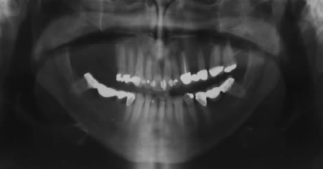
Figure 2. This is a panoramic radiograph taken in May 04, one week after surgery. Note the partial resection of the tuberosity, extraction of first and second molars.
References
- Shear M. Odontogenic keratocysts: natural history and immunohistochemistry. Oral Maxillofacial Surg Clin N Am. 2003; 15: 347-362.
- Oda D, Rivera V et al. Odontogenic keratocyst: the northwestern USA experience. J Contemp Dent Pract. 2000 Feb 15; 1(2): 60-74.
- Zachariades N, Papanicolaou S, Triantafyllou D. Odontogenic keratocysts: Review of the literature and report of sixteen cases. J Oral Maxillofac Surg. 1985; 43: 177-182
- Reichart PA, Philipsen HP, Sonner S. Ameloblastoma: biological profile of 3677 cases. Eur J Cancer B Oral Oncol 1995;31B:86–99.
- Adekeye EO, McLavery K. Recurrent ameloblastoma of the maxillofacial region. Clinical features and treatment. J Maxillofac Surg 1986;14:153-157.
- Gardner DG. Some current concepts on the pathology of ameloblastomas. J Oral Maxillofac Surg 1996;82:660-669.
- Omandi BI, Guthua SW, Awange DO et al. Maxillary obturator prosthesis rehabilitation following maxillectomy for ameloblastoma: case series of five patients. Int J Prosthodont. 2004;17:464-468.
- Whitaker SB, Vigneswaran N, Budnick SD, Waldron CA. Giant cell lesions of the jaws: evaluation of nucleolar organizer regions of varying behavior. J Oral Pathol Med 1993; 22:402-405.
- Tallan EM, Olsen KD, McCaffrey TV, Unni KK, Lund BA. Advanced giant cell granuloma: a twenty-year study. Otolaryngol Head Neck Surg 1994; 110:413-418.
- Carlos R, Sedano HO. Intralesional corticosteroids as an alternative treatment for central giant cell granuloma. Oral Surg Oral Med Oral Pathol 2002; 93:161-166.
- Yamazaki H, Ota Y et al. Brown tumor of the maxilla and mandible: progressive mandibular brown tumor after removal of parathyroid adenoma. J Oral Maxillofac Surg. 2003; 61:719-722.
Sorry! you are incorrect
Although central giant cell granuloma is included on the differential diagnosis, it is the opinion of this author that the radiographic and clinical presentation of this case are highly unlikely to represent CGCG. To be complete, we will describe CGCG. It is a non-neoplastic process that can, but not commonly, behave in a very aggressive and expansile manner, destroying bone and displacing teeth. Over 60% of CGCG cases occur in patients younger than 30 years of age, with twice as many occurrences in females as in males. CGCG is classified into aggressive and non-aggressive types; the aggressive type tends to occur in younger patients and is known to cause disfiguration, especially after surgery. Over 70% of cases occur in the mandible anterior to the first molar tooth. This lesion has also been described in other cranio-facial and small long bones such as those of the hands and feet (8, 9).
The usual treatment for CGCG is surgery, ranging from curettage and en bloc to resection (9). The latter is used in aggressive and recurring cases (9). Treatments alternatives to surgery have emerged with successful results ranging from steroid injections (10) to calcitonin injections or nasal spray to interferon alfa-2a injections (10), which are administered 2-3 times per week for several months. The latter is the most expensive alternative treatment. Neither the histology or the clinical presentation were supportive of CGCG.
Sorry! you are incorrect
This would most likely represent a primary type of hyperparathyroidism. Primary hyperparathyroidism is usually due to an adenoma of the parathyroid gland, but is sometimes a result of hyperplasia (in about 10% of cases) and is rarely caused by adenocarcinoma. Primary hyperparathyroidism is 3 times more common in females than males and typically occurs in patients in their fifties and older (11). The clinical presentation is characteristically referred to as bones, stones, groans and moans—affecting multiple organs including bones, kidney stones, gastrointestinal system groans, muscles and the central nervous system moans. Bone lesions are painful multiple unilocular and multilocular radiolucencies that affect the fingers and the skull, including the jaw bones. They are called ‘brown tumors’ because of the deep chocolate brown color of the specimens resulting from hemorrhage and hemosiderin pigmentation. Stones are result of hypercalcemia affecting the kidneys and skin. Groans are related to intestinal ulcers and constipation, and moans are related to alteration in the central nervous system such as depression and sometimes seizures. This patient did not have alteration in his calcium/phosphate levels (11). Neither the histology or other clinical findings were supportive of brown tumor of hyperparathyroidism.
Sorry! you are incorrect
The destructive bony presentation seen in this case (Fig 1) lends itself to including a malignant neoplasm such as salivary gland neoplasm in the differential diagnosis. The posterior hard palate, anterior and posterior soft palate are ideal locations for malignant salivary gland neoplasms such as adenoid cystic carcinoma, mucoepidermoid carcinoma and polymorphous low grade adenocarcinoma. Although the latter does not readily invade bone, the former two do. These neoplasms usually present as swellings in the soft tissue of the palate, at times however, they may be hidden and grow inwardly into the palate and maxillary sinus rather than the usual outward, visible swelling. We also need to emphasize that although, the palate is the most common oral location for these neoplasms, malignant salivary gland neoplasms can also arise within the sinus salivary gland tissue.
Salivary gland neoplasms in general favor adults with equal sex distribution (sometimes favoring females). They are slow-growing and asymptomatic. Adenoid cystic carcinoma and polymorphous low grade adenocarcinoma tend to invade nerve, therefore leading to clinical symptoms ranging from pain to anesthesia. Nerve invasion is characteristically a sign of advanced disease. Neither, the histology or the clinical presentation of this case are supportive of a salivary gland neoplasm.