Large unilocular radiolucency associated with impacted tooth #17
Can you make the correct diagnosis?
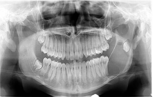
This is a 16-year-old male who was referred by his general dentist to an oral surgeon for a left mandibular swelling.
1. Dentigerous cyst
2. Odontogenic keratocyst
3. Unicystic ameloblastoma
Sorry, you are incorrect!
A radiographic presentation of a well-circumscribed and unilocular radiolucency associated with an impacted tooth should make dentigerous cyst (DC) first on the differential diagnosis list. The age of the patient, the site and the association with impacted third molar tooth are all consistent with that of the typical clinical presentation of a DC. The expansion is also consistent with this cyst but not the bony perforation. The size of this lesion is large but fits the range of sizes for this cyst. The histology however is not consistent with dentigerous cyst.
Congratulations, you are correct!
The radiographic presentation of a well-circumscribed and unilocular radiolucency is also consistent with that of odontogenic keratocyst (OKC), an aggressive cyst of odontogenic origin. The site, posterior mandible extending into the ramus, is also consistent with the clinical presentation of OKC. The association with an impacted tooth is not unusual. The age of this patient falls into the main cluster of occurrence at 10-40 years of age, but OKCs can occur at any age. Bone perforation is part of what characterizes OKC as an aggressive cyst. The expansion is not consistent with the clinical behavior of OKC, which usually grows anterior-posterior rather than expanding in buccal and lingual areas. Larger OKCs can be expansile. In this case, the expansion is mild, which is consistent with the clinical behavior of larger OKCs. The histology is that of odontogenic keratocyst.
Sorry, you are incorrect!
The age, site, association with an impacted tooth, and the unilocular and well-circumscribed radiolucency are all clinical characteristics of unicystic ameloblastoma. Almost 90% of UAs present looking like a dentigerous cyst in the posterior mandible associated with impacted third molar teeth. Unicystic ameloblastomas tend to be very expansile and may perforate bone. The expansion in this case is not prominent, but it is present, and therefore UA should be present on the differential diagnosis. The histology, however, is not consistent with unicystic ameloblastoma.