All cases are discussed by: Dr. Dolphine Oda, UW-Oral Pathology Biopsy Service
Single large gingival swelling left maxilla; area of retained root of tooth #13
Contributed by:
Drs. Andrea Burke & Eleanor Chen
Departments of Oral Surgery & Pathology, UW-Seattle, WA
Case Summary and Diagnostic Information
This patient is a 62-year-old white female who presented with a very large gingival swelling.
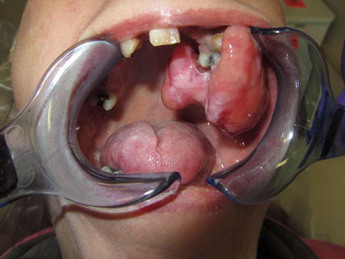
This patient is a 62-year-old white female who presented with a very large gingival swelling. She stated that the swelling had been of about three months’ duration, but she was not certain. The gingival swelling was in the area of retained roots of tooth #13 (Figure 1) measuring 4.2 x 3.4 x 3.7 cm in greatest dimensions. The patient reported that this swelling impaired her ability to close her mouth and impacted eating but that she was otherwise asymptomatic. There was no pain, bleeding or sensitivity associated with the swelling.
Figure 1 This photograph is taken at the first clinical presentation. The figure shows a large smooth surface, pink, nonulcerated, sessile swelling measuring 4.2 X 3.4 X 3.7 cm in greatest dimensions. It is associated with retained root of tooth #13.
The patient’s past medical history is significant for two strokes as well as cigarette and marijuana use.
The patient indicates that this gingival swelling was present for at least three months. It was slow growing but was otherwise asymptomatic. It interfered with eating and closing of the mouth. and was not painful. It did however affect eating and closing of her mouth. This lesion was pink with intact epithelium and was firm on palpation. It was associated with retained root of tooth #13.
The incisional biopsy was performed under local anesthesia while the excisional surgery was performed under general anesthesia. The gingival swelling was completely excised in one piece (Figures 2 & 3). The patient also successfully underwent complete extraction of her teeth and retained roots (Figure 4).
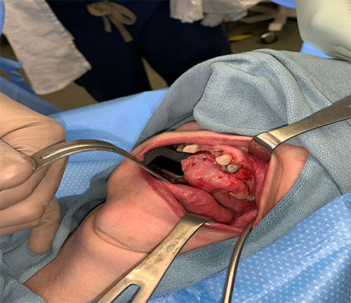
Figure 2 This photograph is taken at the excisional surgery while the patient is under general anesthesia. Note the swelling that being removed in one piece.
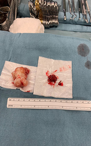
Figure 3 This is the image of the gross specimen as it is completely excised in one piece.

Figure 4 This is a postoperative image of the maxillary alveolar ridge completely healed.
Histologic examination a large and multisected piece of soft tissue composed of keratinized surface epithelium with an underlying mass of moderately cellular fibrous connective tissue containing calcified viable bone (Figures 5-7). The connective tissue mass ranged from fibrotic to moderately cellular. The bony trabeculae were mostly reparative and lamellar in type with viable osteocytes.
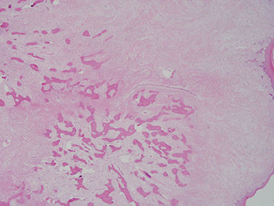
Figure 5 Low power (X40) H & E stained section revealing a low-power image of a moderately cellular mass of connective tissue covered by stratified squamous epithelium. This mass contains bony trabeculae.
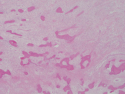
Figure 6 : Higher power (X100) H & E stained section revealing moderately cellular connective tissue mass and bony trabeculae with viable osteocytes. The bony trabeculae are both lamellar and reparative in type.
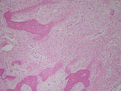
Figure 7 Higher power (X200) H & E stained section revealing a closer look at the connective tissue mass with reparative bony trabeculae.
After you have finished reviewing the available diagnostic information