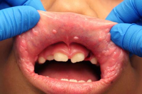
May 2009: Multiple small nodule; lips and bilateral buccal mucosa
Can you make the correct diagnosis?
This is an 8-year-old Hispanic male who was seen at the University of Washington Oral Surgery Clinic. He presented with a chief complaint of multiple small nodules on the upper and lower lips, bilateral buccal mucosa and tongue.
Sorry! you are incorrect
It is valid to consider MHS in this case since almost 80% of patients with this condition have oral lesions which manifest in small nodules on the dorsal tongue, gingival and buccal mucosa. However, almost all patients also have similar skin lesions around the lips, nose and ears. This patient had no skin lesions and the histology was not supportive of MHS.
MHS is a rare disease complex involving the skin, breast, gastrointestinal tract and thyroid. It is autosomal dominant with high penetrance. The gene (PTEN) is mapped to chromosome 10q23 (1-2). The skin lesions are papillomatous, smooth-surfaced nodules and occur in about 99% of patients with this condition, mostly on the face and around the eyelids, nose, mouth and ears in particular. They may also affect the arms and hands. The nodules are mostly trichilemmomas of hair follicle origin. Breast diseases include fibrocystic disease and some malignant neoplasms. Thyroid lesions include goiter and follicular adenocarcinoma. They may also have colon polyps which are typically benign in behavior.
Sorry! you are incorrect
The age of the patient, the location and the multiplicity in this case suggest a virally induced condition; common warts should be seriously considered in such cases. However, the histology is not supportive of multiple verruca vulgaris.
Verruca vulgaris, more commonly known as the “common wart,” is a communicable disease. It is most common in children. In the US, about 22% of children have common warts. This is also a benign lesion induced by human papilloma virus (HPV) types 2 and 4 most commonly but occasionally HPV types 1, 3, 27, 29 and 57 have been reported as well (3-4). They are more commonly of the skin, especially the fingers, than any other location, including the mouth. The oral lesions can be single but are usually multiple and tend to be exophytic, papillary, white and contained or discrete. Oral common warts are usually the result of auto-inoculation, i.e. finger and mouth if the child sucks on his or her finger. They are more common on the lips (vermillion border and labial mucosa) and anterior tongue but are described in other areas including the soft palate and buccal mucosa. Histologically, the oral verruca vulgaris has a distinct verrucous configuration with elongated projections covered by a thick keratin layer, alternating parakeratin and orthokeratin, under which lies a granular cell layer with keratohyalin granules and a thick and confluent spinous layer. Koilocytes are frequently noted in the upper spinous layer and granular cell layer. Treatment includes simple excision for the isolated lesions. For multiple lesions, laser surgery and/or topical antiviral agents are indicated [is this right?]. Up to 30% of common warts tend to spontaneously regress and disappear within 6 months; up to 75% will disappear in three years (5-6). They rarely recur but can be re-infected. To prevent re-infection, the source of infection, i.e. finger lesions, should also be treated.
Sorry! you are incorrect
MEN2b syndrome is also a very important condition to consider in this case. This syndrome presents with multiple small smooth-surfaced nodules on the lower lip and anterior tongue. However, MEN2b patients have marfanoid features (tall, slender and lower jaw prognathism) which this patients does not have. The histology is also not supportive of MEN2b syndrome.
These are rare disease groups affecting the endocrine system. Three types have been described. Some are inherited as autosomal dominant while others develop as a result of mutation. MEN syndrome type 2b is the most significant type to dental practitioners (11-13). The gene for this type has been mapped to chromosome 20p12.2. Lesions appear as early as infancy, causing problems with feeding and normal thriving. During the first decade, multiple small mucosal nodules occur in the oral cavity on the anterior tongue, lower lip, and bilateral corner of mouth. These nodules are highly characteristic of the disease. They may also occur on the eyelids and conjunctiva, and represent multiple neuromas which are histologically made up of hyperplastic peripheral nerve fibers. Multiple melanotic skin lesions have been described in these patients. Patients have marfanoid features, a thick lower lip, and an everted upper eyelid. They also develop pheochromocytoma, profuse sweating, diarrhea, and severe hypertension. In addition, they often develop medullary carcinoma of the thyroid at around 18-25 years of age, but this aggressive neoplasm has been described in patients as young as 23 months. MEN 2b patients demonstrate high levels of catecholamines and calcitonin if pheochromocytoma and medullary carcinoma are present. Preventive removal of the thyroid gland is recommended.
Congratulations! You are correct
Focal epithelial hyperplasia, also known as Heck’s disease, is a rare disease in white and black populations but is common in Native Americans (7-8), especially in the South American Indian population; in this case, the patient is from Guatemala. It is an infectious disease caused by human papilloma virus types 13 and 32. It was first described by Archard et al in the Eskimo population of Greenland (7-10). This condition has a distinct presentation; it occurs in children living in poor conditions and presents in multiple small (around 5mm or slightly larger), slightly elevated, smooth-surfaced and dome-shaped nodules, similar in color to the surrounding mucosa. These lesions can be isolated or coalesced, forming more diffuse and ill-defined elevation of the mucosa. Lip and buccal mucosa are the most common locations, but can also occur on the gingival, palate and other areas. It has been described in adult AIDS patients as multiple papillary lesions similar to focal epithelial hyperplasia. Some are also HPV 13 and 32 positive. Histologically, this condition presents as a blunt dome-shaped epithelial hyperplasia with mitosoids (the latter are not always present). Treatment ranges from doing nothing to laser surgery to topical or injectable chemotherapy. This condition tends to be more persistent in AIDS patients and can be resistant to treatment. In otherwise healthy children, some regress spontaneously and others respond to liquid nitrogen or laser treatment. Other forms of therapy include intra-lesional injections or topical chemotherapy.
References
- Gorlin R, Cohen MM et al. Syndromes of the Head and Neck. 1990 Oxford University Press. Pages 357-360.
- Tsou HC, Ping XL et al. The genetic basis of Cowden’s syndrome: three novel mutations PTEN/MMAC1/TEP1. Human Genet. 1998; 102: 467-473
- Summersgill KF, Smith EM, Levy BT, et al. Human papillomavirus in the oral cavities of children and adolescents. Oral Surg Oral Med Oral Pathol Oral Radiol Endod. Jan 2001;91(1):62-9.
- Gibbs S, Harvey I, Sterling JC, Stark R. Local treatments for cutaneous warts. Cochrane Database Syst Rev. 2003;CD001781.
- Silverberg N. Human papillomavirus infections in children. Curr Opin Pediatr. 2004 Aug;16(4):402-9.
- Silverberg N. Warts and molluscum in children. Adv Dermatol. 2004;20:23-73.
- van der Voort EA, Arani SF, Hegt VN, van Praag MC. Focal epithelial hyperplasia of the oral mucosa. A unique manifestation of human papillomavirus. Ned Tijdschr Tandheelkd. 2009 Mar;116(3):149-51.
- Carlos R, Sedano HO. Multifocal papilloma virus epithelial hyperplasia. Oral Surg Oral Med Oral Pathol. 1994 Jun;77(6):631-5.
- González LV, Gaviria AM, Sanclemente G, Rady P, Tyring SK, Carlos R, Correa LA, Sanchez GI. Clinical, histopathological and virological findings in patients with focal epithelial hyperplasia from Colombia. Int J Dermatol. 2005 Apr;44(4):274-9.
- Bennett LK, Hinshaw M. Heck’s disease: diagnosis and susceptibility. Pediatr Dermatol. 2009 Jan-Feb;26(1):87-9.
- Oda, D. Soft-tissue lesions in children. Oral Maxillofac Surg Clin North Am. 2005 Nov;17(4):383-402.
- Guinto ER, Closmann JJ. Dental rehabilitation of the patient with multiple endocrine neoplasia Type 2b. Gen Dent. 2007 Sep-Oct;55(5):429-35.
- Lips CJM, Hoppener JWM et al. Medullary thyroid carcinoma: role of genetic testing and calcitonin measurement. Annals clinical Biochem. 2001; 38: 168-179.
