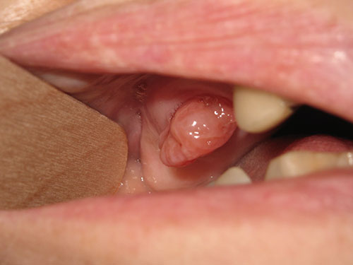
Mass emerging from socket of extracted tooth-right posterior maxilla
Can you make the correct diagnosis?
This is a 73-year-old female with dementia who was presented to a general dentist in October 2019 with a swelling and purulence drainage involving the right posterior maxillary teeth #s 2, 3, 4, and 5 which were extracted.
Sorry, you are incorrect!
EG is a soft tissue lesion that fills an extraction site that failed to heal. This patient’s history is consistent with challenging extraction sites that started with significant infection and draining pus making the area less likely to completely heal even with antibiotics. The clinical presentation of one such socket is not unusual in that scenario. The radiographic changes of a large and completely filled maxillary sinus is not typical of EG. The histology was not consistent with EG.
Sorry, you are incorrect!
The most likely primary cancer emerging from an extraction site would be a misdiagnosed gingival squamous cell carcinoma (SCC) mistaken for severe periodontitis especially in heavy smokers and heavy alcohol consumers. This patient has no history of smoking which in and by itself would not exclude primary gingival squamous cancer emerging from the extraction socket. The age of this is consistent SCC but not the gender. Poor oral hygiene can contribute to SCC of the gingiva which this patient had. The histology was not consistent with gingival SCC
As to metastatic cancer to the jaw, it is not unusual for carcinomas especially from breast cancer and lung cancer patients to metastasize to the jaw. Posterior mandible is the most common site for cancer metastasis, not the maxilla. Although this patient has no history of cancer, in 30% of cases, metastasis to the oral cavity is the first presentation. The histology was not consistent with a malignant neoplasm.
Sorry, you are incorrect!
The site and the age of this patient are not typical for solid ameloblastoma. Gender is fine since ameloblastoma occurs equally in both males and females. The age is on the older age range of 30-70 years of age. Mean age however is around 33 years of age. The site is not consistent since over 80% of ameloblastomas occur in the posterior mandible and 15% occur in the maxilla. The lesion filling the maxillary since is consistent with ameloblastoma behavior that occur in this site where they tend to fill the maxillary sinus before showing the typical clinical swelling. The histology was not consistent with ameloblastoma
Congratulations, you are correct!
This patient presented with significant amount of pus draining from multiple teeth and from her right nostril. Chances of extraction leading to oro-antral fistula is high. Therefore a number of benign and malignant sinonasal diseases can herniate through that socket. The histology was that of Oncocytic Schneiderian Papilloma (OSP). The histology for this case benign epithelial and showed no evidence of in situ or invasive malignancy.
Schneiderian papillomas arise from the Schneiderian membrane. These papillomas are not common and are benign but can develop in situ or invasive malignancy such as squamous cell carcinoma or other carcinomas. If we have 100 Schneiderian papillomas of the sinonasal site, only 3% are those of the Oncocytic type which makes it especially rare. The first clinical presentation through an extraction socket is unique as well.