Left mandible, lingual-exophytic radiopaque lesion
Can you make the correct diagnosis?
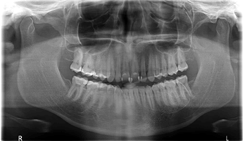
This is a 28-year-old Caucasian male who presented with an exophytic lingual plate expansion in the left posterior mandible.
Sorry, you are incorrect!
Torus mandibularis is a lingual mandibular bony growth located above the mylohyoid ridge, the age of the patient, and the history of slow growth of the lesion over the last 12-15 years, combined with the radiographic findings of a radiopaque exophytic nodule with no capsule, are all consistent with torus mandibularis. However, a unilateral position is unusual for this condition. Torus mandibularis usually presents in a bilateral manner but not symmetrical. The histology is not consistent with torus mandibularis.
Sorry, you are incorrect!
The radiopaque, well-demarcated exophytic nodule protruding from the mandible are all characteristics consistent with osteomas. The patient’s age and gender are also consistent with osteomas, since they occur at an average age of 30 and twice as often in males as in females. The mandible is a more common location for osteomas of the jaws, and the radiographic appearance of a protruding bony nodule with no capsule is also consistent with that condition. The clinical history of the lesion having been present since the patient’s teenage years, with a slow growth over 12 to 15 years, is unusual for an osteoma of this size. There is a variant of osteoma known to grow slowly over many years, but it tends to be very large (much larger than this lesion), thus the given name of “giant osteoma.” The histology is not consistent with osteoma.
Sorry, you are incorrect!
The history of the lesion growing for 12 to 15 years and the onset of disease during teenage years is consistent with fibrous dysplasia (FD). The continued slow growth to the current age is also somewhat consistent with FD since it usually completes growth by age 30. The exophytic swelling and the radiopaque presentation lacking a radiolucent rim are also consistent with the diagnosis of FD. However the well-demarcated nature of this lesion is not consistent with FD since FD tends to blend in with the surrounding normal bone. The site of the patient’s lesion is also not consistent with FD, which more commonly occurs in the maxilla. The histology is not consistent with FD.
Sorry, you are incorrect!
The exophytic swelling protruding from the posterior mandible lingually is consistent with a clinical presentation of central ossifying fibroma (COF). Almost 90% of COFs occur in the premolar-molar area of the mandible. This patient’s age is compatible with the average age of 35 years for COF. However, COF are rare in males. They occur around five times more commonly in females than in males. The radiographic findings for this patient are also not consistent with COF, since the latter presents as mixed radiolucent/radiopaque lesion with a radiolucent rim. The histology is also not consistent with COF.
Congratulations, you are correct!
Intraosseous hemangiomas of the jaw (mandible and maxilla) are exceedingly rare but when they occur, they most commonly occur in the mandible (3:1 mandible over maxilla) and particularly in the body of the mandible. This is consistent with this patient’s presentation. The age of this patient is consistent with intraosseous hemangioma of the jaw according to the oral pathology literature which reports that the jaw lesions occur in the first three decades of life. However, the literature related to other bone hemangiomas i.e. vertebrae and skull bone indicates that more patients are in the fifth decade of life with a wide age range of 2-85. These lesions tend to be slightly more common in females with a female to male ratio of 3:2 so the gender in this case is not typical of intraosseous hemangiomas. The radiographic finding of a protruding exophytic radiopaque lesion is especially rare for intraosseous hemangiomas in general including those of the jaw. Radiopaque and expansile intraosseous hemangiomas are previously reported but simulating fibrous dysplasia. More typically, intra-osseous hemangiomas are radiolucent with a unilocular, multilocular, or soap-bubble morphology, and occasionally with a sun-ray appearance. They can occur in, or involve, the inferior alveolar canal, rendering it larger and irregular. The slow growth of this patient’s lesion over a 12 to 15 year period is unusual for hemangiomas in general. The soft tissue hemangioma tend to grow fast for the first one to two years and slow or stop growing thereafter and may even regress after puberty. The growth pattern of the bone counterpart is not clearly defined. The histology of this patient’s specimen is consistent with cavernous hemangioma.