Single Dome-Shaped Swelling on the Hard Palate
Can you make the correct diagnosis?
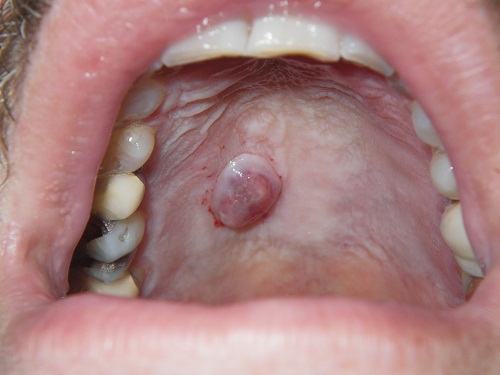
This is a 51-year-old male who presented with a smooth-surfaced, focally ulcerated, grey-blue, firm nodule on the hard palate.
Sorry! you are incorrect
A swelling in the middle of the hard palate should bring to mind torus palatinus, but not in this case. There are many reasons for this, the most important being that the consistency of this swelling is firm but not as hard as bone. Tori and exostoses would have a bony hard consistency that this swelling lacked. So torus palatinus is off the list of the differential diagnosis. The differential diagnosis for a firm soft tissue swelling would include a salivary gland neoplasm. The location is good for that diagnosis and the color is more in favor of a mucoepidermoid carcinoma. The histology, however, is not supportive of this diagnosis.
Mucoepidermoid carcinoma is a malignant neoplasm of salivary gland origin that can present as a smooth-surfaced swelling or a non-healing ulcer on the palate, usually the posterior lateral palate. It occurs in a wide age range and has three histologic types: low, intermediate and high; the low-grade type is more common in the oral cavity. Mucoepidermoid carcinoma accounts for 10% of all salivary gland neoplasms. While the majority of MECs occur in the parotid gland, some also occur in minor salivary glands, especially the palate, tongue, buccal mucosa, lips, and retromolar pad areas. It can occur at any age with a predilection for occurrence in young people. Studies by the Armed Forces Institute of Pathology (AFIP) find 44% of cases occurring in patients under 20 years of age, most commonly on the palate. Their youngest patient was nine months old. The low-grade lesions are slow-growing and painless, and not encapsulated; they sometimes resemble a mucocele, especially those at the retromolar pad area. Mucoceles of the retromolar pad area are rare, and for that reason it is best to biopsy them early to exclude the possibility of a mucoepidermoid carcinoma masquerading as a mucocele. High-grade lesions tend to be more common in the parotid gland; they present as rapidly growing, painful lesions with facial nerve paralysis and sometimes with regional lymph node metastasis. Histologically, mucoepidermoid carcinomas consist of a variety of cell types and architectural patterns which constitute the three histologic gradings. Although low-grade mucoepidermoid carcinoma is characterized by an abundance of mucous-producing cells and duct-like structures with cystic dilation, the mere presence of certain types of cells and architecture should not be used to determine the histologic grade. Complete surgical removal with clean margins is the preferred treatment for the low-grade type. Radiation therapy has also been successfully used, especially when the tumor involves the surgical margins.
Sorry! you are incorrect
A soft tissue swelling in the palate should bring to mind salivary gland neoplasms but are usually present in the posterior/lateral palate, rather than middle palate as is the case here. The most common salivary gland neoplasm of the major and minor glands is pleomorphic adenoma, which should therefore be on the differential diagnosis. The color in this case, however, is not supportive of PA. The age of the patient fits the description of PA, but not the gender. The histology is not supportive of this diagnosis.
Pleomorphic adenoma is the most common benign salivary gland neoplasm of both the major and minor salivary glands. It originates from the myoepithelial cells and the reserve cells of the intercalated ducts. It accounts for 80% of all benign salivary gland neoplasms. It occurs in both major and minor salivary glands and accounts for up to 77% of parotid, 68% of submandibular, and 43% of minor salivary gland tumors. It is most common in females 30-50 years of age, but it is also rarely described in children. One study reports 1% of cases affecting children under 10 years of age and 5.9% between the ages of 10-20. It presents as a small, painless, slowly enlarging nodule. If left untreated, it can enlarge significantly, sometimes increasing by several pounds in weight. It occurs in the oral cavity, especially the palate and lips. On the palate, it is usually located in the posterior hard palate or anterior soft palate but can also be in the posterior soft palate; PA usually occurs in the posterior and lateral palate, as opposed to torus palatinus, which usually occurs in the middle hard palate and in the anterior. The posterior hard palate mixed tumor is fixed due to the bone-bound anatomy of the region; the tumor is otherwise movable. Histologically, mixed tumor has a wide variety of cellular and pattern manifestations; the main cellular components are epithelial duct-like structures and mesenchymal-like tissue such as myxochondroid matrix. These lesions are generally encapsulated, ranging from predominantly myxoid (36%) to extremely cellular (12%). Complete surgical removal with clean margins is the preferred treatment. Palatal lesions respond well to excision in one piece with the periosteum and overlying mucosa. Pleomorphic adenoma has a good prognosis, but it has a tendency for recurrence (up to 44%) if not treated thoroughly. The risk of recurrence is less if it occurs in the minor salivary glands (up to 20%). The risk of malignant transformation is about 5%.
Sorry! you are incorrect
A firm, well-demarcated nodule in the hard palate should make a clinician consider a lesion of peripheral nerve origin, especially palisaded encapsulated neuroma. The age of this patient is indicative of this lesion, but the color and the size are not. The histology is not supportive of palisaded encapsulated neuroma.
Palisaded encapsulated neuroma, also known as solitary circumscribed neuroma, is a benign solitary lesion of peripheral nerve origin, believed to be a reactive rather than neoplastic process. Some suggest that it is reactive to trauma. It was first described on the skin in 1972 and in the oral cavity in 1976. The head and neck area is the most common location for this condition. It occurs most often in the mouth, particularly on the hard palate. It usually presents as an isolated lesion, though it may occur in multiples on rare occasions. It is a small (less than one cm in size), exophytic, smooth surfaced and pink (same color as the surrounding mucosa) nodule. It is usually painless and can be of a long duration. Mucosal sites that are affected include the nose, mouth and glans penis. Palisaded encapsulated neuroma occurs in adults between the fifth and seventh decade with no gender predilection. Conservative surgical removal is the treatment of choice.
Congratulations! You are correct
A firm soft tissue nodule of the palate with grey-blue color should make a clinician think of leiomyoma or angiomyoma (vascular leiomyoma). The age and gender in this case is supportive of leiomyoma. The histology is also supportive of leiomyoma.
Leiomyoma is a benign neoplasm of smooth muscle origin. Uncommon in the oral cavity, it mainly occurs in the G.I. tract, uterus and skin. In the oral cavity, it most likely originates from the vascular smooth muscle. Vascular leiomyoma, also known as angiomyoma, accounts for 75% of oral leiomyomas. Patients tend to be over 30 years of age and are predominantly males. It is most common on the posterior tongue, palate, cheeks and lips. It is a slow growing, painless, pedunculated, smooth-surfaced, normal color or slightly bluish nodule. Intra-osseous leiomyomas have been reported, but very rarely. Histopathologically, this neoplasm is made up of a well-circumscribed to encapsulated nodule composed of bundles of smooth muscle cells, with some surrounding blood vessels in cases of angiomyoma. Simple and conservative excision is the treatment of choice.
References
- Auclair PL, Ellis GL. Mucoepidermoid carcinoma. In Ellis GL, Auclair PL, Gnepp DR, editors. Surgical pathology of the salivary glands. Philadelphia: W.B. Saunders, 1991. p. 269-298.
- Hicks J, Flaitz C. Mucoepidermoid carcinoma of salivary glands in children and adolescents: assessment of proliferation markers. Oral Oncol. 2000 Sep;36(5):454-60.
- Brandwein MS, Ivanov K, Wallace DI, Hille JJ, Wang B, Fahmy A, Bodian C, Urken ML, Gnepp DR, Huvos A, Lumerman H, Mills SE. Mucoepidermoid carcinoma: a clinicopathologic study of 80 patients with special reference to histological grading. Am J Surg Pathol 2001; 25:835-45.
- Pons Vicente O, Almendros Marqués N, Berini Aytés L, Gay Escoda C. Minor salivary gland tumors: A clinicopathological study of 18 cases. Med Oral Patol Oral Cir Bucal. 2008 Sep 1;13(9):E582-8.
- Toida M, Shimokawa K, Makita H, Kato K, Kobayashi A, Kusunoki Y, Hatakeyama D, Fujitsuka H, Yamashita T, Shibata T. Intraoral minor salivary gland tumors: a clinicopathological study of 82 cases. Int J Oral Maxillofac Surg. 2005 Jul;34(5):528-32. Epub 2005 Jan 24.
- Yih WY, Kratochvil FJ, Stewart JC. Intraoral minor salivary gland neoplasms: review of 213 cases. J Oral Maxillofac Surg. 2005 Jun;63(6):805-10.
- Hamakawa H, Takarada M, Ito C, Tanioka H. Bone-forming pleomorphic adenoma of the upper lip: report of a case. J Oral Maxillofac Surg. 1997 Dec;55(12):1471-5.
- Koutlas IG, Scheithauer BW. Palisaded encapsulated (“solitary circumscribed”) neuroma of the oral cavity: a review of 55 cases. Head Neck Pathol. 2010 Mar;4(1):15-26. doi: 10.1007/s12105-010-0162-x. Epub 2010 Jan 23.
- Jordan RC, Regezi JA. Oral spindle cell neoplasms: a review of 307 cases. Oral Surg Oral Med Oral Pathol Oral Radiol Endod. 2003;95:717–724. doi: 10.1067/moe.2003.140.
- Tomich CE, Moll MC. Palisaded encapsulated neuroma of the lip. J Oral Surg. 1976;34:265–268.
- Anastassov GE, van Damme PA. Angioleiomyoma of the upper lip: report of a case. Int J Oral Maxillofac Surg. 1995 Aug;24(4):301-2.
- Maeda Y, Hirota J, Osaki T, Hayashi K, Sonobe H, Otsuki Y. Angiomyoma of the upper lip: report of a case with electron microscopic and immunohistochemical observation. Br J Oral Maxillofac Surg. 1989 Jun;27(3):236-42.
- Gianluca S, Marini R, Tonoli F, Cristalli MP. Leiomyoma of oral cavity: case report and literature review. Ann Stomatol (Roma). 2011 Jan;2(1-2):9-12.