Expansile, Radiopaque/Radiolucent Lesion, Right Posterior Mandible
Can you make the correct diagnosis?
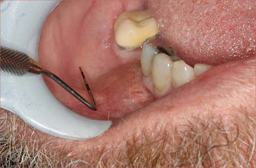
The patient is a 68-year-old male who was referred to the oral surgeon with persistent swelling at the extraction site of tooth #30.
Sorry! you are incorrect
Given the clinical presentation of four months of swelling and discomfort in an extraction site, together with the radiographic findings of a predominantly radiopaque/focally radiolucent lesion, chronic sclerosing osteomyelitis would be a reasonable condition to include on the differential diagnosis, even possibly as the first diagnosis to consider. However, the histology of this specimen was not supportive of osteomyelitis.
Chronic sclerosing osteomyelitis is a challenging condition to diagnose both clinically and histologically. This condition has gone by a series of names, starting with “ossifying osteomyelitis”, a term used by Thoma in the 1940s. The current name was first used by Shafer in 1957 where he divided the histopathological patterns into “focal” and “diffuse” subtypes. This condition is most commonly found in the mandible, is more prevalent in females, and usually presents with pain and local swelling with periodic exacerbations. Most investigators agree that this type of osteomyelitis is not usually associated with suppuration or fistula formation. Some reports indicate that mild suppuration may develop, but this occurs rarely.
Chronic sclerosing osteomyelitis can occur at any age, including in children under the age of ten. The type that occurs in children is called “juvenile mandibular chronic osteomyelitis” which affects 6-12 year old children, mostly girls; it occurs mainly in the mandible. The adult type affects people 30-40 years of age, and is twice as common in females but shows no race predilection. It too is by far more common in the mandible, especially the posterior mandible, as is the case in this patient. Patients, children or adults, present with a history of years of pain with episodic exacerbations. Some investigators have been successful in isolating E corrodens from the lesions, while most have not been able to grow any micro-organisms. Radiographically, chronic sclerosing osteomyelitis presents as diffuse, irregular aggregates of bone with indistinct borders. It may mimic Paget’s disease. Histologically, the biopsy shows dense bone with a “mosaic” consistent with a reactive process. The surrounding soft tissue may contain neutrophils during the acute stage, and variable number of chronic inflammatory cells, mainly lymphocytes and plasma cells. It is indeed a challenge to treat chronic sclerosing osteomyelitis. Antibiotics, especially penicillin and doxycycline, and decortications seem to be effective at the early stages may help prevent the disease from progression. Resection has occasionally and in rare cases, been used. The literature indicates that NSAIDS are useful in controlling the intensifying pain. Hyperbaric oxygen is reported to be effective in some cases.
Sorry! you are incorrect
The age and gender of this patient, the radiographic findings and the persistence of the disease should make one think of a hard tissue producing primary malignant neoplasm such as chondrosarcoma. The histology here, however, is not supportive of chondrosarcoma.
Chondrosarcoma is a malignant mesenchymal neoplasm of cartilage. It is uncommon, especially in the jaws. It occurs more often in the maxilla, particularly in the incisor area. It can occur at any age but is most common in males in the sixth decade of life. This neoplasm has a low tendency for metastasis. In general, the prognosis is better than that of osteosarcoma, but can vary depending on the stage of the disease. It commonly presents as an asymptomatic swelling with buccal and lingual expansion. Patients may experience unexplained paresthesia, headache, loosening and loss of teeth. Radiographically, it presents as an ill-defined, mottled radiolucency with snowflake or punctate calcifications or diffuse radiopacity as is the case in this patient. Sometimes, the teeth will show a widened periodontal ligament space. Histologically, it is characterized by immature and pleomorphic cartilage, but at times the cartilage is benign-looking. It is rare for the cartilage to calcify or differentiate and form bone. Therapy for chondrosarcoma ranges from wide local excision to radical resection with or without chemotherapy, depending on the stage of disease. The prognosis ranges from good to poor.
Sorry! you are incorrect
The predominant radiopaque radiographic appearance in the above case should make one think of fibrous dysplasia. However, the age of the patient, the location of the lesion, and the history of the condition developing shortly after a tooth was extracted are not supportive of the clinical behavior of FD; neither is the histology.
Fibrous dysplasia is a tumor-like developmental and hamartomatous disorder of bone that presents in three forms: monostotic, polyostotic and craniofacial. It is common, comprising 7% of all benign bone tumors. The monostotic form constitutes approximately 80% of all fibrous dysplasia cases. It affects males and females equally, occurs in childhood and adolescence, and usually completes growth by the age of 30. The craniofacial form affects the maxilla and the craniofacial complex, and is more aggressive than the monostotic form. Polyostotic cases constitute 10% of all FDs; they may (such as McCune-Albright) or may not (Jaffe) be associated with multiple endocrine disorders.
The etiology of FD is not clear but a mutation (GNAS 1) has been identified at multiple chromosomes. This mutation can occur at all ages, even in infancy. Some of the FD cases are clinically asymptomatic and are found incidentally; many present with swelling and deformity, and rare cases involve pain. Swelling of the maxilla or mandible is the most common presentation. It may involve bones other than the maxilla, including the zygoma, sphenoid and frontal. Monostotic FD is usually unilateral and is known to displace the surrounding teeth, but is otherwise firmly seated. It is usually slow-growing, but rapid growth has been described, especially during puberty. With rapid growth, temporary pain is also described.
The radiographic appearance, especially of the maxilla, is classically described as a ground glass appearance where fine radiopacity is noted. The mandibular lesions are much more deceiving; more variability exists, thus making the radiographic diagnosis more difficult. In the mandible, the appearance ranges from cystic radiolucency to a classical ground glass radio-opacity. Histologically, it consists of vascular fibrous connective tissue stroma with irregular (Chinese character) spicules of woven bone. Giant cells may be seen. At times, artifactual shrinkage of the connective tissue from bone is noted. Many cases do not need treatment; those that do are best treated with surgical recontouring of the affected bone to address aesthetic or functional concerns. This is preferably performed after cessation of growth due to the high incidence of requirement for secondary procedures. Some sources report a tendency for increased growth of the lesion following surgical intervention, but the evidence for this is weak. Even if recontouring is performed in adulthood, multiple procedures are frequently required. This may be due to inadequate removal, continued growth or the ossification of the subperiosteal hematoma, which is difficult to avoid due to the very vascular nature of the dysplastic bone. Radical excision with primary reconstruction of the affected bone has been suggested, but is not widely accepted. Radiation therapy has been used in the past and is successful in stopping growth of the lesion. Unfortunately, a significant incidence of development of osteosarcoma in the irradiated bone prohibits the use of this modality. Bisphosphonates have successfully been used in controlling FD growth, especially the large lesions. The overall prognosis is good with proper clinical management.
Congratulations! You are correct
This patient had no history of primary prostate cancer when tooth #30 was extracted. It was weeks after the extraction of tooth #30 that a lytic lesion of the lower spine was identified and biopsied, resulting in a diagnosis of metastatic prostate cancer.
Cancer metastasis to the oral cavity is neither specific nor common. Although it constitutes less than 1% of all oral malignant neoplasms, it may have devastating implications for the patient, mainly because metastasis to other sites has already developed or is inevitable. Theoretically, any malignant neoplasm can metastasize to the oral cavity, but in actuality few do and out of the ones that do, the majority are carcinomas rather than sarcomas. The malignant neoplasms that metastasize most commonly to the mouth are primary cancers of the breast, lung, kidney and prostate. The only cancer that commonly produces bone-containing metastases in the jaws is prostate cancer, although occasionally breast cancer can produce bone as well. The metastatic lesions from primary cancers arising in other sites are usually radiolucent. Metastatic neoplasms from the prostate are sometimes misdiagnosed as benign fibro-osseous lesions.
Lung and prostate cancers are the most common neoplasms to metastasize to the oral cavity in men, but malignant neoplasms of the thyroid, pancreas, colon, and liver may also do so. By far the most common location for cancer metastasis to the mouth is the posterior mandible, where 80% of cases occur, followed by the gingiva. The maxilla is a rare location for tumor metastasis, described mainly in adults over the age of 30 and rarely in children. Pain and swelling are the most common clinical symptoms, and were seen with this patient. Oral metastases may also present asymptomatically, simulate a periapical lesion, cause palatal or gingival swelling like a pyogenic granuloma, or cause anesthesia and parasthesia, especially when it involves the inferior alveolar canal. (The latter circumstance results in so-called “numb-chin syndrome.” ) Tooth loosening, displacement and sharp resorption have also been described and occurred in this patient with 3+ mobility of tooth # 31 associated with discomfort. The radiographic appearance of irregular sclerotic bone formation is common for prostate, and occasionally for breast, cancers that metastasize to the mandible. The majority of the metastatic neoplasms cause bony destruction with ill-defined borders, the moth-eaten appearance of some bony destruction indicating aggressive behavior. It is also important to mention that at times, well-demarcated, but not corticated, lesions with a benign morphology have also been described.
As in this case, the diagnosis of tumor metastasis to the oral cavity carries a poor prognosis because the oral cavity is usually not an isolated site of metastasis and generally indicates more disseminated tumor. Patients are typically treated with chemotherapy and the five-year survival rate is unfortunately very low.
References
- WG Shafer, Chronic sclerosing osteomyelitis, Oral Surg. 1957:15; 138-142.
- Marx RE, Carlson ER, Smith BR, Toraya N. Isolation of Actinomyces species and Eikenella corrodens from patients with chronic diffuse sclerosing osteomyelitis. J Oral Maxillofac Surg. 1994;52(1):26-34.
- Y. Suei, K. Tanimoto, M. Miyauchi and T. Ishikawa, Partial resection of the mandible for the treatment of diffuse sclerosing osteomyelitis: Report of four cases, J Oral Maxillofac Surg. 1997: 55; 410.
- MacDonald-Jankowski DS. Fibro-osseous lesions of the face and jaws. Clin Radiol. 2004;59:11-25.
- Rao VV, Schnittger S et al. G protein Gs alpha (GNAS 1), the probable candidate gene for Albright hereditary osteodystrophy, is assigned to human chromosome 20q12-q13.2. Genomics. 1991;10:257-261.
- Neville BW, Damm DD, Allen CM, Bouquot JE. Oral & Maxillofacial Pathology. 3rd ed. Philadelphia: WB Saunders Company 2009.
- Anil S, Beena VT et al. Chondrosarcoma of the maxilla. Case report. Aust Dent J. 1998;43:172-174.
- Hayt MW, Becker L et al. Chondrosarcoma of the maxilla: panoramic radiographic and computed tomographic with multiplanar reconstruction findings. Dentomaxillofac Radiol. 1998;27:113-116. A.
- Hirshberg and A. Buchner, Metastatic tumours to the oral region. An overview. Oral Oncol. 1995; 31: 355–360.
- Ivan der Waal, RIF, Buter, J. Oral metastases: report of 24 cases. Br J Oral Maxillofac Surg. 2003; 41: 3-6.
- Neville BW, Damm DD, Allen CM, Bouquot JE. Metastasis to the oral soft tissue. In: Oral and Maxillofacial Pathology, 3rd edition. Philadelphia: W.B. Saunders, 2007.
- Ogunyemi O, Rojas A, Hematpour K, Rogers D, Head C, Bennett C. Metastasis of genitourinary tumors to the head and neck region. Eur Arch Otorhinolaryngol. 2009.