Multilocular Radiolucent/Radiopaque Lesion, Anterior Mandible
Dolphine Oda, BDS, MSc
doda@u.washington.edu
Contributed by Dr. Brian Rubens
Oral & Maxillofacial Surgery, Bothell, WA
Case Summary and Diagnostic Information
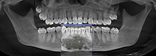
This is a 17-year-old female with expanding anterior mandible. A panoramic view of the anterior mandible shows a large and slightly well-demarcated multilocular radiolucent lesion interspersed with foci of mixed radiolucent/radiopaque component.
Diagnostic Information Available
This is a 17-year-old female with expanding anterior mandible. A panoramic view of the anterior mandible shows a large and slightly well-demarcated multilocular radiolucent lesion interspersed with foci of mixed radiolucent/radiopaque component (Figures 1 & 2). This lesion is of unknown duration and the swelling is described to measure 5.0 x 1.5 x 2.0 cm at its greatest dimensions. The swelling is noted more in the buccal vestibule. It is otherwise asymptomatic and the associated teeth are vital.
Figure 1. This radiograph was taken at the patient’s first clinical presentation. Note the relatively well-demarcated and mostly multilocular radiolucent lesion with foci of mixed radiolucent and radiopaque lesion in the anterior mandible crossing the midline.
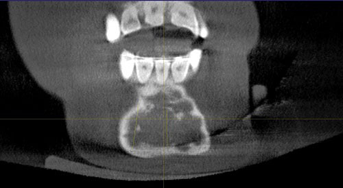
Figure 2. This radiograph was taken in first clinical presentation. Note the relatively well-demarcated and mostly multilocular radiolucent lesion in the anterior mandible crossing the midline.
The patient’s past medical history is unremarkable.
The patient reported mild swelling of the anterior buccal mandible (Figures 1 & 2). It is of unknown duration and is asymptomatic. Clinical examination revealed slight expansion of the anterior buccal vestibule. All the anterior teeth tested vital.
Figure 1. This radiograph was taken at the patient’s first clinical presentation. Note the relatively well-demarcated and mostly multilocular radiolucent lesion with foci of mixed radiolucent and radiopaque lesion in the anterior mandible crossing the midline.
Figure 2. This radiograph was taken in first clinical presentation. Note the relatively well-demarcated and mostly multilocular radiolucent lesion in the anterior mandible crossing the midline.
Treatment
Under intravenous anesthetic an incision was made over an intact buccal cortex. An incisional biopsy was performed and the tissue submitted for microscopic evaluation.
Excisional Biopsy
Histologic examination reveals multiple pieces of decalcified hard and soft tissue composed of a fibro-osseous lesion (Figure 3). The specimen is made up of cellular fibrous connective tissue stroma intermixed with calcified bone. The latter is at various stages of development and the trabeculae are of variable shapes and sizes (Figure 4). Osteoblastic rimming is absent in most parts (Figure 5). In many areas, the bony trabeculae show evidence of artifactual retraction from the connective tissue stroma (Figures 3 & 4).
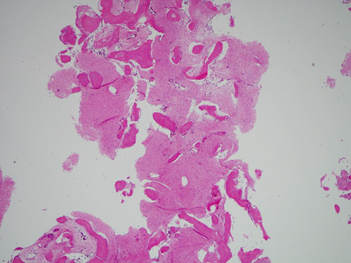
Figure 3. Low power (x40) H & E histology shows a benign fibro-osseous lesion which is made up of strands of spindle-shaped fibroblasts focally forming bone. The bony trabeculae show evidence of artifactual retraction from the connective tissue stroma.
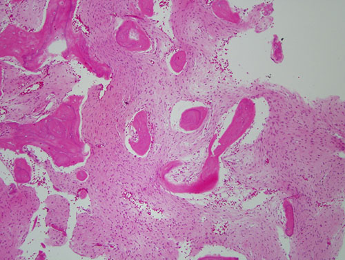
Figure 4. Higher power (x100) H & E histology shows a closer view of the benign fibro-osseous lesion which is made up of strands of spindle-shaped fibroblasts focally forming bone. The bony trabeculae show evidence of artifactual retraction from the connective tissue stroma. Also note the absence of osteoblastic rimming around the bony trabeculae.
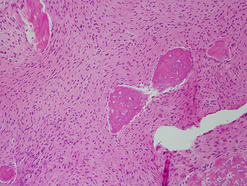
Figure 5. Higher power (x200) H & E histology shows strands of spindle shaped cells surrounding bony trabeculae lacking osteoblastic rimming.
After you have finished reviewing the available diagnostic information