Return to Case of the Month Archives
Multilocular Expansile Radiolucency, Posterior Mandible
Dolphine Oda, BDS, MSc
doda@u.washington.edu
Contributed by
Drs. Tony Rea, Ross Beirne and Andrew Tolas
Seattle, Washington
Case Summary and Diagnostic Information
This is a 26-year-old male who is a recent immigrant from Mexico. He presented for the evaluation of a right mandibular swelling of two years’ duration which has become progressively larger during that time.
Diagnostic Information Available
This is a 26-year-old male who is a recent immigrant from Mexico. He presented for the evaluation of a right mandibular swelling of two years’ duration which has become progressively larger during that time. It is otherwise asymptomatic, with no pain, numbness or enlarged lymph nodes
His past medical and family history is unremarkable. The patient is in excellent health and is on no medications. He has no history of tobacco use, but occasionally drinks alcohol.
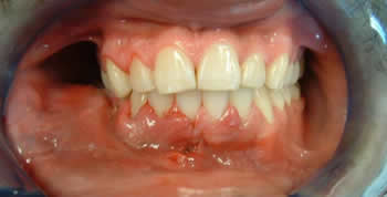
The patient presented with palpable buccal expansion of the right posterior mandible. The expansion measured ~3cm X 5cm and extended from tooth #27-#32. The expansion was visible clinically (Fig 1). There was no evidence of pain or altered sensation on the lower lip or chin. A large cavity was noted on tooth #30. The floor of mouth was soft and not elevated. There was mild lingual expansion in the area of tooth #32. Radiographic changes demonstrated a well-defined multilocular radiolucency (Fig 2) with a “honeycomb” pattern. There is also evidence of tooth displacement, tooth resorption and inferior alveolar nerve displacement (Fig 2).
Figure 1. This is a clinical view of the lesion at presentation demonstrating obliteration of the right mandibular vestibule indicating an expansile swelling in the right mandible.
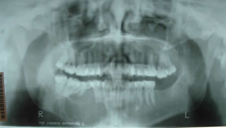
Figure 2. This is a panoramic radiograph demonstrating a well-demarcated and multilocular radiolucent lesion between teeth #s 27-32, slightly displacing teeth.
The specimen revealed fragments of loose and myxoid fibrous connective tissue (Fig 4). It is made up of stellate fibroblasts suspended on a delicate network of collagen fibrils and small blood vessels (Fig 5).
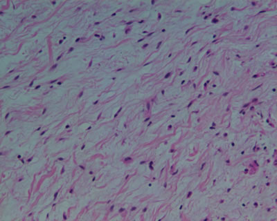
Figure 4. H & E stained section at 20X magnification (low scanning) demonstrating a mass of lamellar bone covered by a thin mucosa. The surface epithelium is stretched and lacks filiform papillae.
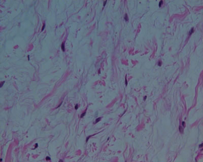
Figure 5. Higher power (x200) histology illustrates a few spindle-shaped fibroblasts suspended on a delicate network of collagen fibers.
After you have finished reviewing the available diagnostic information