Unilocular radiolucency right ramus
Can you make the correct diagnosis?
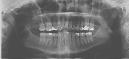
This is a 44-year-old Caucasian female who presented to the clinic for black spot on the jaw x-ray.
Sorry, you are incorrect!
This patient had no symptoms at all: no swelling, no pain and no numbness. This was an incidental finding. It is unusual for unicystic ameloblastoma to present with no symptoms at all. At the very least, it is expansile. The unilocular and well-demarcated radiolucency is consistent with the radiographic presentation of UA but the site being only in the ramus without involving the posterior mandible (a tooth-bearing area) is also unusual for UA. The lack of association with an impacted third molar is not consistent with UAs since almost 90% of these neoplasms are associated with an impacted tooth simulating a dentigerous cyst. The age of this patient is old for UA; they usually occur in patients under 20 years of age. For these reasons, UA should be taken off the differential diagnosis list. The histology is also not consistent with UA.
Sorry, you are incorrect!
The lack of clinical symptoms is not unusual for OKC because these cysts/tumors are not expansile or painful. They grow anterior-posteriorly, hollowing the jaw. They usually occur in the posterior mandible/ramus area and appear on radiographs as a well-demarcated unilocular radiolucency. Like unicystic ameloblastoma, it is unusual for OKC to occur in the ramus alone without involving the posterior mandible (the tooth-bearing area). The age of this patient is consistent with OKC, which can occur at any age, but is most common between 20 and 40 years of age. Therefore, this condition should remain on the differential diagnosis. The histology, however, is not consistent with OKC.
Sorry, you are incorrect!
While this patient’s gender is consistent with central (intraosseous) hemangioma, the age is not. It is rare for central hemangiomas to be asymptomatic and found incidentally, but this kind of presentation has been reported. The radiographic findings and the site in the ramus area anterior to the inferior alveolar canal are also reported in rare hemangiomas of the jaws. Therefore, this condition should be included on the differential diagnosis.
Intraosseous or central hemangiomas are usually symptomatic, typically presenting with symptoms such as mild expansion, pain, paresthesia, pulsation or, occasionally, localized gingival bleeding on pressure. On rare occasions, they can be asymptomatic. Unlike the soft tissue hemangiomas, intraosseous or central hemangiomas are rare conditions anywhere in the body, including the jaws. They occur more commonly in the vertebrae and skull bone. When they occur in the jaws, the mandible is affected twice as often as the maxilla. Within the mandible, they occur most often in body of the mandible, followed by the ramus in the inferior alveolar canal area, which is consistent with this patient’s clinical presentation. They are typically slow-growing and are usually radiolucent. The radiolucency may be unilocular or may have a soap-bubble or spider-web appearance. Occasionally, they have a sun-ray appearance. In case of the presence of Phleboliths, calcifications can also be identified. Clinically, they are most common in the first two decades of life and occur in females by a ratio of 2:1. They can resorb and displace teeth and can cause bleeding around the involved teeth. The histology in this case is not consistent with intraosseous hemangioma.
Congratulations, you are correct!
Peripheral nerve tumors (in this case schwannoma)—Intraosseous
The age, gender, site, and radiographic presentation of a well-demarcated unilocular radiolucency anterior to the inferior alveolar canal are all consistent with intraosseous (central) peripheral nerve tumors of the jaws. However, the incidental finding of an asymptomatic condition is unusual in the case of intraosseous peripheral nerve tumors, since most such tumors of the jaws are symptomatic. In one study, three of 35 cases were asymptomatic; therefore, this condition should remain on the differential diagnosis.
Intraosseous peripheral nerve tumors occur in a wide age range of 2 to 72 has been reported, with an average age of 29. It is twice as common in females as in males. A clinical presentation of expansion is the most common sign of this condition, followed by pain and burning sensation. It occurs most often in the posterior mandible followed by the ramus involving the inferior alveolar canal. The radiographic presentation is typically a well-demarcated or corticated radiolucency, either unilocular or multilocular. The histology in this case was consistent with a benign peripheral nerve tumor with specific features of schwannoma.