Mixed radiolucent radiopaque lesion associated with impacted tooth #32
Can you make the correct diagnosis?
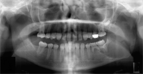
This is a 15-year-old white female who presented with a history of increasing swelling without pain in the left mandible. Her mother noted the swelling and took her to the family dentist for a clinical evaluation where a periapical radiograph was taken. The dentist then referred this patient to Dr. Grace who took a panoramic radiograph (Fig 1).
Sorry! you are incorrect
The scalloped radiolucency, the location and age of the patient are consistent with the radiographic and clinical presentation of an OKC. However, significant swelling (expansion) is not consistent with OKC. The histology in this case was not that of an OKC.
The Odontogenic keratocyst is an aggressive odontogenic cyst, known for its rapid growth ad its tendency to invade the adjacent tissues, including bone. It has a high recurrence rate and is associated with basal cell nevus syndrome. It affects patients in the age ranges of 20-29 and 40-59, but cases in patients ranging in age from 5 to 80 years have been reported (1). The distribution between sexes varies from equal distribution to a male-to-female ratio of 1.6:1, except in children. Odontogenic keratocysts may occur in any part of the upper and lower jaw, with the majority (almost 70%) occurring in the mandible. They occur most commonly in the angle of the mandible and ramus (2). Radiographically, OKCs present predominantly as unilocular radiolucencies with well-defined or sclerotic borders; they may also present as multilocular radiolucencies, but rarely. OKCs commonly present as unilocular radiolucency with scalloped borders. Teeth associated with OKC are vital. OKCs grow to sizes larger than any other odontogenic cysts. They usually penetrate the bone rather than expand it and grow in an anterior to posterior direction (1, 2). Despite this aggressive growth, they often remain asymptomatic, thus growing to large sizes and hollowing the bone. Treatment of choice is surgery with cauterization especially with Carnoy’s solution.
Sorry! you are incorrect
The expansion, the location and the age of the patient are all in support of a unicystic ameloblastoma. However, 90% of unicystic ameloblastomas occur in association with an impacted third molar especially mandibular third molar. This patient does not have a third molar and the radiolucency is not associated with any impacted tooth. This may also be the radiographic and clinical presentation of a solid or multilocular ameloblastoma since the radiograph has a hint of multilocular appearance. The age of the patient however is young for the solid or multilocular follicular type ameloblastoma. The histology was not supportive of unicystic or follicular ameloblastoma.
Ameloblastoma is one of the most common benign neoplasms of odontogenic origin. It accounts for 11% of all odontogenic neoplasms/hamartomas (3-4). It is a slow-growing, persistent, and locally aggressive neoplasm of epithelial origin. It affects a wide range of age distribution but is mostly a disease of adults, at an average age of 33, with equal sex distribution. Reports from Africa and India show a male predilection; it also has a predilection for occurrence in black patients (5). The location and age of this patient can be consistent with an ameloblastoma. The unilocular radiolucency is unusual for the solid type but is consistent with the 10% of the unicystic ameloblastomas that occur in an extra-follicular manner. About 85% of ameloblastomas occur in the posterior mandible; most of these occur in the molar-ramus area, and some occur in the anterior mandible. Three types are described. Solid ameloblastoma is characteristically expansile, radiolucent and multilocular and not consistent with this case. The unicystic type is radiographically unilocular but in 90% of the time is associated with the crown of an impacted tooth (4-5). The other 10% are not and can present between teeth as is the case here and for that reason, it should be considered on the differential diagnosis. Ameloblastoma, if not treated, can reach very large sizes, causing facial disfigurement. It loosens, displaces and resorbs adjacent teeth. Ameloblastomas are usually not painful unless infected, in which case it can be mildly painful. Parasthesia and anesthesia are extremely rare, unless the lesion is very large in size. Also, ameloblastoma tends to expand rather than perforate the cortical bone; if the latter occurs with extension into the adjacent soft tissue, it has a higher tendency for recurrence and therefore a worse prognosis than cases in which the ameloblastoma is completely encased by bone (3-5). The solid type is treated with en bloc or resection with clean margins. Curettage is the treatment of choice for the unicystic type. The histology of this case is not supportive of ameloblastoma.
Congratulations! You are correct
The jawbones are subject to many cysts, mostly odontogenic (of tooth) origin, some developmental (of both tooth and non-tooth origin), while others are cyst-like or pseudocyst structures such as the salivary gland depression and traumatic bone cavity. Traumatic bone cyst is best called traumatic bone cavity since this condition does not represent a true cyst. Traumatic bone cavity (TBC) is not unique to the jawbones; it is also described in the long bones and is known as a simple solitary bone cyst occurring mostly in the humerus or femur, close to the epiphyseal plate (6). The long bone simple cyst is similar to the jaw traumatic bone cavity radiographically and occurs in the same age range. Trauma has been suggested as the etiology along with other non-substantiated theories such as cystic degeneration of a preexisting tumor or of the fatty marrow in the area.
Some reports suggest that it is more common in males (7) while others report equal distribution between males and females (6). The long bone counterpart is more common in males by a ratio of 2.5:1. Most reports agree that the average age of occurrence is below 20 years of age (6-7). These lesions can occur, but are uncommon, over the age of 30. Kaugars reported a higher number of traumatic bone cavity in African American females compared to the literature (6). The latter patients were over the age of 30 (6). This may suggest an association with florid cemento-osseous dysplasia. The mandible is the most commonly affected area, where over 95% of the cases occur, especially in the posterior premolar-molar area. They rarely extend to the ramus; therefore this case is unique in that it extends very high into the ramus. Also, TBC are known to cross the midline anteriorly. In one study, 27% were anterior to the canine and some crossed the midline. They are usually unilocular and radiolucent, typically above the alveolar canal and in many cases with a scalloped superior border spreading between the roots of teeth. The latter are vital and are frequently found hanging within the empty cavity. About 25% of the lesions present in the anterior mandible apical to the canine tooth and usually are round and unilocular, which can be mistaken for a periapical lesion leading to an unnecessary endodontic treatment. Therefore, it is important to test the vitality of the teeth and carefully examine the radiographs for changes consistent with a periapical granuloma or cyst. Large, expansile and multilocular traumatic bone cavities (as is the case in this patient) have been described, but are rare. Expansion is not characteristic of TBC but it is described in about 26% of the cases (6). They are otherwise asymptomatic. The margins of these lesions range from very well defined to corticated to punched out radiolucency. Pathologic fractures associated with traumatic bone cavity have been described in the jaws, but are rare. They are however more common with those of the long bones.
Clinically, surgeons report an empty cavity at entrance in about two thirds of the cases and straw-colored fluid filled cavities in about one third of the cases. Blood clot is also present occasionally. The bony cavity is scraped to generate bleeding, which is considered the treatment of choice for this condition. Other methods of treatment have been tried, such as packing the curetted cavity with autogenous blood, autogenous bone and hydroxyapatite (7). Various other reports demonstrate healing of traumatic bone cavity after injection of autogenous blood, after aspiration and after endodontic treatment. These lesions may spontaneously heal, but rarely. Biopsy material consists of fragments of viable bone and loose connective tissue, as reported in our case. Osteoclast-like giant cells have also been described in a few cases (6). Exploration surgery usually leads to healing. Recurrence is rare.
Treatment
Under IV sedation, the area was surgically explored. The surgeon found minimal fluid and a cavity with no cystic lining. The cavity was quite large and the encasing cortical bone showed evidence of perforation in multiple areas. The cavity was debrided aggressively to generate bleeding and subsequent clotting that would stimulate bone regeneration. Small fragments of hard and soft tissue were submitted for microscopic examination.
References
- Shear M. Odontogenic keratocysts: natural history and immunohistochemistry. Oral Maxillofacial Surg Clin N Am. 2003; 15: 347-362.
- Oda D, Rivera V et al. Odontogenic keratocyst: the northwestern USA experience. J Contemp Dent Pract. 2000 Feb 15; 1(2): 60-74.
- Reichart PA, Philipsen HP, Sonner S. Ameloblastoma: biological profile of 3677 cases. Eur J Cancer B Oral Oncol 1995;31B:86–99.
- Gardner DG. Some current concepts on the pathology of ameloblastomas. J Oral Maxillofac Surg 1996;82:660-669.
- Omandi BI, Guthua SW, Awange DO et al. Maxillary obturator prosthesis rehabilitation following maxillectomy for ameloblastoma: case series of five patients. Int J Prosthodont. 2004;17:464-468
- Kaugars GE, Cale AE. Traumatic bone cyst. Oral Surg Oral Med Oral Pathol Oral Radiol Endod 1987; 63: 318-324.
- Dellinger TM, Holder R et al. Alternative treatments for a traumatic bone cyst: a longitudinal case report. Quintessence Int. 1998; 29: 497-502.