Return to Case of the Month Archives
Ulcerated Swelling in the Hard Palate
Dolphine Oda, BDS, MSc
doda@u.washington.edu
Contributed by
Dr. Roman Carlos and Dr. Elisa Contreras
Centro de Medicina Oral /University Mariano Galvez, Guatemala
Case Summary and Diagnostic Information

This is a 57-year-old Guatemalan female who complained of a palatal swelling that she first noticed eight months prior to the clinical consultation. The swelling was firm, ulcerated and asymptomatic.
Diagnostic Information Available
This is a 57-year-old Guatemalan female who complained of a palatal swelling that she first noticed eight months prior to the clinical consultation. The swelling was firm, ulcerated and asymptomatic.
Her past medical history is unremarkable. She underwent a cesarean section procedure for the birth of her only son 36 years ago but has not had any other noteworthy illnesses or surgeries. At the time of the consultation, she was not on any medications and had not had a dental procedure performed in recent months.
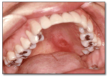
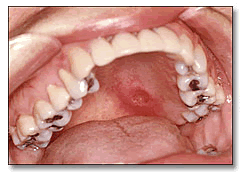
Clinical examination revealed a firm swelling in the posterior and lateral hard palate (Fig 1) bordering on the anterior soft palate. The swelling was ulcerated; however, the patient did not recall having traumatized the area. She reported that the lesion had steadily increased in size over the past eight months. An incisional biopsy was performed.

Figure 1.
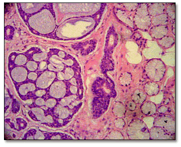
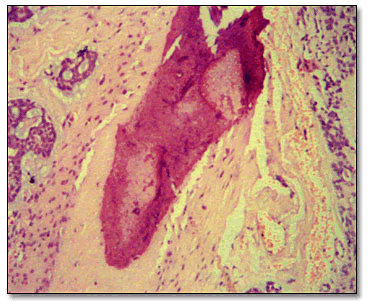
Histological examination revealed an infiltrative salivary gland neoplasm of epithelial origin. It is made up of uniform and basaloid cells with hyperchromatic nuclei. These cells are arranged in nests and lobules with a cribriform pattern (Fig 2). The neoplastic cells show no evidence of pleomorphism or mitosis and yet they are infiltrative of the surrounding connective tissue, salivary gland lobules and bone (Fig 3). The morphology of the surgical specimen was similar to that of the incisional biopsy.

Figure 2.

Figure 3.
After you have finished reviewing the available diagnostic information