Multiple Papillary Lesions, Generalized, Oral Cavity
Can you make the correct diagnosis?
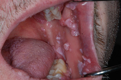
This is a 44-year-old white male with numerous pink/white papillary lesions, some of which are exophytic and many of which are flat and spongy.
Sorry! you are incorrect
Almost 80% of patients with multiple hamartoma syndrome have oral lesions manifested in numerous small nodules on the dorsal tongue, gingiva and buccal mucosa. However, almost all of these patients also have similar skin lesions around the lips, nose and ears. This patient had no skin lesions other than in the perianal area. In addition, MHS nodules occur at the end of the second or third decade of life, whereas our patient is 44 years of age and these lesions are of two years’ duration. The histology is not supportive of MHS.
MHS is a rare disease complex involving the skin, breast, gastrointestinal tract and thyroid. It is autosomal dominant with high penetrance. The gene (PTEN) is mapped to chromosome 10q23 (1-2). The skin lesions are papillomatous, smooth-surfaced nodules and occur in about 99% of patients with this condition, mostly on the face and around the eyelids, nose, mouth and ears in particular. They may also affect the arms and hands. The nodules are mostly trichilemmomas of hair follicle origin. Breast diseases include fibrocystic disease and some malignant neoplasms. Thyroid lesions include goiter and follicular adenocarcinoma. Patients may also have colon polyps; these are typically benign in behavior.
Congratulations! You are correct
Condyloma acuminatum consists of benign and papillomatous proliferations of surface epithelium induced by HPV types 6 and 11, and sometimes types 16 and 18 (the latter are more common in genital condylomas) (4-6). It is transmitted via contact, and most cases are sexually transmitted. It accounts for 20-30% of all sexually transmitted diseases (4-6). Condyloma is more common in the genitalia but can be transmitted to the mouth via oral-genital contact; in this case, all lesions should be treated, as should the patient’s sexual partner(s). It usually presents as multiple, membranous pink and papillomatous lesions but can also present as a single pinkish or white lesion. In the mouth, the commissures and dorsal tongue are more commonly affected, but it can occur anywhere else including the gingiva, floor of mouth and upper lip. Oral and peri-anal condylomas, as well as other HPV-type papillary lesions including common warts and focal epithelial hyperplasia, become very aggressive, generalized and proliferative in immune-comprised individuals (such as in AIDS patients, which is the case with this patient) (3-8). Histologically, condylomas present with broad papillary projections with cells demonstrating viral changes such as empty cytoplasm (koilocytes) and meiosis. The connective tissue is loose and vascular and is infiltrated by lymphocytes. The treatment of choice for condylomas in the general population ranges from chemical cauterization or laser treatment to conventional surgery in the case of a single lesion. The key to proper treatment is to first treat the genital lesions before treating the oral lesions to prevent autoinoculation. It is also very important that the patient’s sexual partner(s) also be treated to prevent transmission between them and the patient; otherwise, the lesions will continue to occur as a result of re-inoculation. In AIDS patients, other modalities are used, such as 5FU cream for genital lesions and alpha interferon injections for oral lesions.
No content for this section. Make sure you wrap your content like this:
Content here
- Gorlin R, Cohen MM et al. Syndromes of the Head and Neck. 1990 Oxford University Press. Pages 357-360.
- Tsou HC, Ping XL et al. The genetic basis of Cowden’s syndrome: three novel mutations PTEN/MMAC1/TEP1. Human Genet. 1998; 102: 467-473.
- Summersgill KF, Smith EM, Levy BT, et al. Human papillomavirus in the oral cavities of children and adolescents. Oral Surg Oral Med Oral Pathol Oral Radiol Endod. Jan 2001;91(1):62-9.
- Anderson CA, Boller AM, Richardson CJ, Balcos EG, Zera RT. Anal condyloma: a comparison between HIV positive and negative patients. Am Surg. 2004 Nov;70(11):1014-8.
- Saiag P, Bauhofer A, Bouscarat F, Aquilina C, Ortonne JP, Dupin N, Mougin C. Imiquimod 5% cream for external genital or perianal warts in human immunodeficiency virus-positive patients treated with highly active antiretroviral therapy: an open-label, noncomparative study. Br J Dermatol. 2009 Oct;161(4):904-9. Epub 2009 May 15.
- Aubin F, Pretet JL, Jacquard AC et al. Human papillomavirus genotype distribution in external acuminata condylomata: a Large French National Study (EDiTH IV). Clin Infect Dis 2008; 47:610–15.Gibbs S, Harvey I, Sterling JC, Stark R. Local treatments for cutaneous warts. Cochrane Database Syst Rev. 2003;CD001781.
- Van Der Voort EA, Arani SF, Hegt VN, van Praag MC. Focal epithelial hyperplasia of the oral mucosa. A unique manifestation of human papillomavirus. Ned Tijdschr Tandheelkd. 2009 Mar;116(3):149-51.
- Carlos R, Sedano HO. Multifocal papilloma virus epithelial hyperplasia. Oral Surg Oral Med Oral Pathol. 1994 Jun;77(6):631-5.
- Gorlin R, Cohen MM et al. Syndromes of the Head and Neck. 1990 Oxford University Press. Pages 392-397.
- Bongiorno MR, Pistone G, Aricò M. Manifestations of the tongue in Neurofibromatosis type 1. Oral Dis. 2006 Mar;12(2):125-9.
- Guinto ER, Closmann JJ. Dental rehabilitation of the patient with multiple endocrine neoplasia Type 2b. Gen Dent. 2007 Sep-Oct;55(5):429-35.
- Lips CJM, Hoppener JWM et al. Medullary thyroid carcinoma: role of genetic testing and calcitonin measurement. Annals clinical Biochem. 2001; 38: 168-179.
Sorry! you are incorrect
Multiple cutaneous nodules are described in the majority of patients with neurofibromatosis, while up to 66% will have oral manifestation of multiple nodules. This patient did not have skin lesions other than peri-anal ones, which suggests the condition is not NFI. In addition, NFI occurs early in life, as early as at birth or the first year of life and usually no later than the teenage years. This patient developed these lesions in his forties. The histology is also not supportive of NFI.
Neurofibromatosis type I (NFI) was first described by Frederick von Recklinghausen in 1882 and for that reason has also been known as von Recklinghausen’s Neurofibromatosis. It is a common autosomal-dominant disease affecting 1 in 2,500-3,000 newborns with 50% mutation and almost 100% penetrance (9-10). The genetic mutation of NFI has been mapped to chromosome 17q11.2 (9-10). Nine types have been described; types I and II are well delineated while the other types are rare and not well studied. Type I accounts for more than 90% of cases. This is followed in frequency by type II, also known as acoustic type, in which bilateral acoustic neuromas are described, leading to hearing loss starting as early as the teenage years. Type II is much less common and affects 1 in 40,000 individuals. The genetic mutation of NFII is mapped to chromosome 22 (9).
The main diagnostic features of neurofibromatosis type 1 are cutaneous neurofibromas, café-au-lait spots (six or more of 1.5 cm size and larger), axillary freckling (Crowe’s sign), Lisch nodules, and several other lesions (9-10). The latter include lesions involving the central nervous system such as mental retardation in about 8% of patients, the cardiovascular system such as pulmonic valvular stenosis, the skeletal system such as scoliosis and the endocrine system such as sexual precocity. Lesions also occur in the eyes, such as neurofibromas of the eyelids and Lisch nodules of the iris. Cutaneous neurofibromas occur at birth and are present at puberty in 60% of cases. They increase in number with time and can number from a few to thousands. They can be present on the skin or in internal organs such as the heart, brain or the gastrointestinal system. They can be small and demarcated or large and pendulous.
Oral soft tissue manifestations of neurofibromatosis can occur in 8-66% of cases, and jaw lesions in 58% of patients (9). The most common lesion is neurofibroma, which occurs mostly in the soft tissue, especially the tongue. It may also affect the jaw bones. Widening of the mandibular foramen and inferior alveolar canal are also described. Other lesions include fungiform papillae, hyperplasia of the soft palate and displaced and unerupted teeth.
Histologically, the neurofibromas can be the conventional, more common type of haphazardly arranged neural tissue in a relatively demarcated nodule or can be the plexiform type where nerve tissue is arranged in lobules. The latter histology is pathognomonic of neurofibromatosis. In the head and neck area plexiform neurofibroma can be associated with the trigeminal nerve branches.
Treatment in Neurofibromatosis patients is primarily for esthetic and functional reasons. Scalpel surgery, laser or dermabrasion treatments have been used for the removal of skin neurofibromas. Malignant transformation has been described in about 4-12% of patients, most of which are peripheral nerve-type malignancies that tend to be aggressive. Other malignancies have been described, including leukemia, rhabdomyosarcoma, Wilm’s tumor, and Pheochromocytoma (9). The general dentist may be the first person to make the primary diagnosis of Neurofibromatosis. Therefore, it is important to know of this disease and its characteristic clinical manifestations. These patients should be referred to their physician for further clinical workup, including a genetic workup.
Sorry! you are incorrect
Multiple Endocrine Neoplasia (MEN) 2b Syndrome
MEN2b syndrome should also be considered since it presents with multiple small smooth-surfaced nodules on the lower lip and anterior tongue. However, this patient had nodules and other flat lesions all over the mouth. In addition, MEN2b occurs at infancy or very early in life. The histology in this case is also not supportive of MEN2b syndrome.
MEN syndromes are rare disease groups affecting the endocrine system. Three types have been described. Some are inherited as autosomal dominant while others develop as a result of mutation. MEN syndrome type 2b is the most significant type to dental practitioners (11-12). The gene for this type has been mapped to chromosome 20p12.2. Lesions appear as early as infancy, causing problems with feeding and normal thriving. During the first decade, multiple small mucosal nodules occur in the oral cavity on the anterior tongue, lower lip, and bilateral corner of mouth. These nodules are highly characteristic of the disease. They may also occur on the eyelids and conjunctiva, and represent multiple neuromas which are histologically made up of hyperplastic peripheral nerve fibers. Multiple melanotic skin lesions have been described in these patients. Patients have marfanoid features, a thick lower lip, and an everted upper eyelid. They also develop pheochromocytoma, profuse sweating, diarrhea, and severe hypertension. In addition, they often develop medullary carcinoma of the thyroid at around 18-25 years of age, but this aggressive neoplasm has been described in patients as young as 23 months. MEN 2b patients demonstrate high levels of catecholamines and calcitonin if pheochromocytoma and medullary carcinoma are present. Preventive removal of the thyroid gland is recommended.